6EOP | pdb_00006eop

DPP8 - SLRFLYEG, space group 20


Experimental Data Snapshot

  • Method: X-RAY DIFFRACTION
  • Resolution: 2.40 Å
  • R-Value Free: 
    0.237 (Depositor), 0.238 (DCC) 
  • R-Value Work: 
    0.214 (Depositor), 0.216 (DCC) 
  • R-Value Observed: 
    0.215 (Depositor) 

Starting Model: experimental
View more details

wwPDB Validation 3D Report Full Report

Validation slider image for 6EOP

Ligand Structure Quality Assessment 


This is version 1.4 of the entry. See complete history

Literature

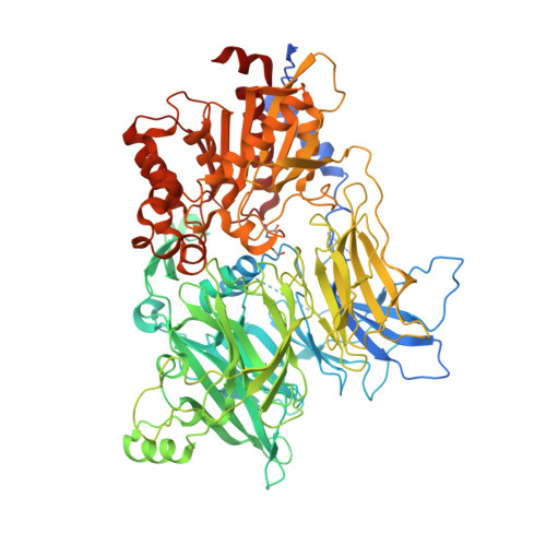
Structures and mechanism of dipeptidyl peptidases 8 and 9, important players in cellular homeostasis and cancer.

Ross, B.Krapp, S.Augustin, M.Kierfersauer, R.Arciniega, M.Geiss-Friedlander, R.Huber, R.

(2018) Proc Natl Acad Sci U S A 115: E1437-E1445

  • DOI: https://doi.org/10.1073/pnas.1717565115
  • Primary Citation Related Structures: 
    6EOO, 6EOP, 6EOQ, 6EOR, 6EOS, 6EOT

  • PubMed Abstract: 

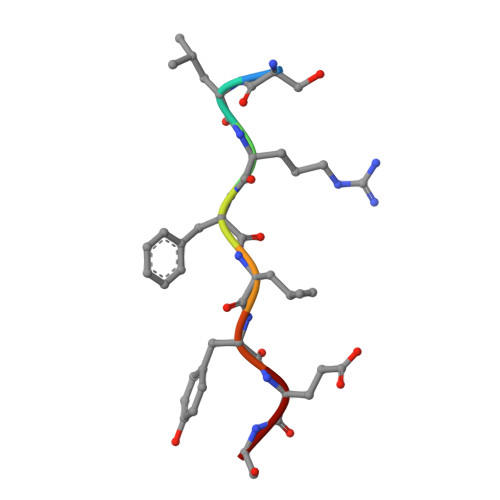
    Dipeptidyl peptidases 8 and 9 are intracellular N-terminal dipeptidyl peptidases (preferentially postproline) associated with pathophysiological roles in immune response and cancer biology. While the DPP family member DPP4 is extensively characterized in molecular terms as a validated therapeutic target of type II diabetes, experimental 3D structures and ligand-/substrate-binding modes of DPP8 and DPP9 have not been reported. In this study we describe crystal and molecular structures of human DPP8 (2.5 Å) and DPP9 (3.0 Å) unliganded and complexed with a noncanonical substrate and a small molecule inhibitor, respectively. Similar to DPP4, DPP8 and DPP9 molecules consist of one β-propeller and α/β hydrolase domain, forming a functional homodimer. However, they differ extensively in the ligand binding site structure. In intriguing contrast to DPP4, where liganded and unliganded forms are closely similar, ligand binding to DPP8/9 induces an extensive rearrangement at the active site through a disorder-order transition of a 26-residue loop segment, which partially folds into an α-helix (R-helix), including R160/133, a key residue for substrate binding. As vestiges of this helix are also seen in one of the copies of the unliganded form, conformational selection may contributes to ligand binding. Molecular dynamics simulations support increased flexibility of the R-helix in the unliganded state. Consistently, enzyme kinetics assays reveal a cooperative allosteric mechanism. DPP8 and DPP9 are closely similar and display few opportunities for targeted ligand design. However, extensive differences from DPP4 provide multiple cues for specific inhibitor design and development of the DPP family members as therapeutic targets or antitargets.


  • Organizational Affiliation
    • Max Planck Institut für Biochemie, D-82152 Martinsried, Germany; ross@biochem.mpg.de huber@biochem.mpg.de.

Macromolecule Content 

  • Total Structure Weight: 314.67 kDa 
  • Atom Count: 21,351 
  • Modeled Residue Count: 2,540 
  • Deposited Residue Count: 2,718 
  • Unique protein chains: 2

Macromolecules

Find similar proteins by:|  3D Structure
Entity ID: 1
MoleculeChains  Sequence LengthOrganismDetailsImage
Dipeptidyl peptidase 8
A, B, C
898Homo sapiensMutation(s): 0 
Gene Names: DPP8DPRP1MSTP097MSTP135MSTP141
EC: 3.4.14.5
UniProt & NIH Common Fund Data Resources
Find proteins for Q6V1X1 (Homo sapiens)
Explore Q6V1X1 
Go to UniProtKB:  Q6V1X1
PHAROS:  Q6V1X1
GTEx:  ENSG00000074603 
Entity Groups
Sequence Clusters30% Identity50% Identity70% Identity90% Identity95% Identity100% Identity
UniProt GroupQ6V1X1
Sequence Annotations
Expand
Reference Sequence
Find similar proteins by:  Sequence   |   3D Structure  
Entity ID: 2
MoleculeChains  Sequence LengthOrganismDetailsImage
SER-LEU-ARG-PHE-LEU-TYR-GLU-GLY
D, E, F
8Homo sapiensMutation(s): 0 
UniProt & NIH Common Fund Data Resources
Find proteins for P63165 (Homo sapiens)
Explore P63165 
Go to UniProtKB:  P63165
PHAROS:  P63165
GTEx:  ENSG00000116030 
Entity Groups
UniProt GroupP63165
Sequence Annotations
Expand
Reference Sequence

Small Molecules

Ligands 3 Unique
IDChains Name / Formula / InChI Key2D Diagram3D Interactions
F15

Query on F15


Download:Ideal Coordinates CCD File
K [auth D],
L [auth E],
M [auth F]
PENTADECANOIC ACID
C15 H30 O2
WQEPLUUGTLDZJY-UHFFFAOYSA-N
FLC

Query on FLC


Download:Ideal Coordinates CCD File
G [auth A]CITRATE ANION
C6 H5 O7
KRKNYBCHXYNGOX-UHFFFAOYSA-K
CA

Query on CA


Download:Ideal Coordinates CCD File
H [auth A],
I [auth B],
J [auth C]
CALCIUM ION
Ca
BHPQYMZQTOCNFJ-UHFFFAOYSA-N
Modified Residues  1 Unique
IDChains TypeFormula2D DiagramParent
CME
Query on CME
A, B, C
L-PEPTIDE LINKINGC5 H11 N O3 S2CYS

Experimental Data & Validation

Experimental Data

  • Method: X-RAY DIFFRACTION
  • Resolution: 2.40 Å
  • R-Value Free:  0.237 (Depositor), 0.238 (DCC) 
  • R-Value Work:  0.214 (Depositor), 0.216 (DCC) 
  • R-Value Observed: 0.215 (Depositor) 
Space Group: C 2 2 21
Unit Cell:
Length ( Å )Angle ( ˚ )
a = 162.83α = 90
b = 246.371β = 90
c = 261.191γ = 90
Software Package:
Software NamePurpose
REFMACrefinement
XDSdata reduction
xia2data scaling
PHASERphasing

Structure Validation

View Full Validation Report


Ligand Structure Quality Assessment 


Entry History 

Deposition Data

Revision History  (Full details and data files)

  • Version 1.0: 2018-02-07
    Type: Initial release
  • Version 1.1: 2018-02-14
    Changes: Database references
  • Version 1.2: 2018-02-28
    Changes: Database references
  • Version 1.3: 2024-01-17
    Changes: Data collection, Database references, Derived calculations, Refinement description
  • Version 1.4: 2024-10-16
    Changes: Structure summary