6EOF

Crystal structure of AMPylated GRP78 in ADP state


Experimental Data Snapshot

  • Method: X-RAY DIFFRACTION
  • Resolution: 1.59 Å
  • R-Value Free: 0.225 
  • R-Value Work: 0.206 
  • R-Value Observed: 0.207 

wwPDB Validation   3D Report Full Report


Ligand Structure Quality Assessment 


This is version 1.1 of the entry. See complete history


Literature

AMPylation targets the rate-limiting step of BiP's ATPase cycle for its functional inactivation.

Preissler, S.Rohland, L.Yan, Y.Chen, R.Read, R.J.Ron, D.

(2017) Elife 6

  • DOI: https://doi.org/10.7554/eLife.29428
  • Primary Citation of Related Structures:  
    5O4P, 6EOB, 6EOC, 6EOE, 6EOF

  • PubMed Abstract: 

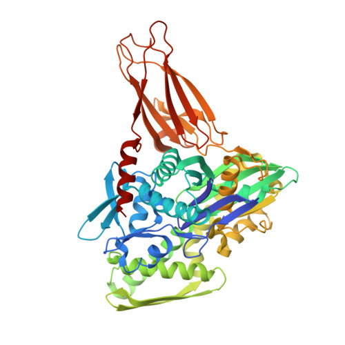
    The endoplasmic reticulum (ER)-localized Hsp70 chaperone BiP contributes to protein folding homeostasis by engaging unfolded client proteins in a process that is tightly coupled to ATP binding and hydrolysis. The inverse correlation between BiP AMPylation and the burden of unfolded ER proteins suggests a post-translational mechanism for adjusting BiP's activity to changing levels of ER stress, but the underlying molecular details are unexplored. We present biochemical and crystallographic studies indicating that irrespective of the identity of the bound nucleotide AMPylation biases BiP towards a conformation normally attained by the ATP-bound chaperone. AMPylation does not affect the interaction between BiP and J-protein co-factors but appears to allosterically impair J protein-stimulated ATP-hydrolysis, resulting in the inability of modified BiP to attain high affinity for its substrates. These findings suggest a molecular mechanism by which AMPylation serves as a switch to inactivate BiP, limiting its interactions with substrates whilst conserving ATP.


  • Organizational Affiliation

    Cambridge Institute for Medical Research, University of Cambridge, Cambridge, United Kingdom.


Macromolecules
Find similar proteins by:  (by identity cutoff)  |  3D Structure
Entity ID: 1
MoleculeChains Sequence LengthOrganismDetailsImage
78 kDa glucose-regulated protein522Cricetulus griseusMutation(s): 1 
Gene Names: I79_019946
UniProt
Find proteins for G3I8R9 (Cricetulus griseus)
Explore G3I8R9 
Go to UniProtKB:  G3I8R9
Entity Groups  
Sequence Clusters30% Identity50% Identity70% Identity90% Identity95% Identity100% Identity
UniProt GroupG3I8R9
Sequence Annotations
Expand
  • Reference Sequence
Experimental Data & Validation

Experimental Data

  • Method: X-RAY DIFFRACTION
  • Resolution: 1.59 Å
  • R-Value Free: 0.225 
  • R-Value Work: 0.206 
  • R-Value Observed: 0.207 
  • Space Group: P 21 21 21
Unit Cell:
Length ( Å )Angle ( ˚ )
a = 69.047α = 90
b = 75.61β = 90
c = 97.5γ = 90
Software Package:
Software NamePurpose
REFMACrefinement
XDSdata reduction
autoPROCdata scaling
PHASERphasing

Structure Validation

View Full Validation Report


Ligand Structure Quality Assessment 


Entry History & Funding Information

Deposition Data


Funding OrganizationLocationGrant Number
Wellcome TrustUnited Kingdom082961/Z/07/Z
Wellcome TrustUnited Kingdom200848/Z/16/Z
British Heart FoundationUnited KingdomPG/12/41/29679

Revision History  (Full details and data files)

  • Version 1.0: 2017-11-01
    Type: Initial release
  • Version 1.1: 2024-01-17
    Changes: Data collection, Database references, Refinement description