6EN6

Crystal structure B of the Angiotensin-1 converting enzyme N-domain in complex with a diprolyl inhibitor.


Experimental Data Snapshot

  • Method: X-RAY DIFFRACTION
  • Resolution: 1.80 Å
  • R-Value Free: 0.226 
  • R-Value Work: 0.193 
  • R-Value Observed: 0.194 

wwPDB Validation   3D Report Full Report


Ligand Structure Quality Assessment 


This is version 2.1 of the entry. See complete history


Literature

The Design and Development of a Potent and Selective Novel Diprolyl Derivative That Binds to the N-Domain of Angiotensin-I Converting Enzyme.

Fienberg, S.Cozier, G.E.Acharya, K.R.Chibale, K.Sturrock, E.D.

(2018) J Med Chem 61: 344-359

  • DOI: https://doi.org/10.1021/acs.jmedchem.7b01478
  • Primary Citation of Related Structures:  
    6EN5, 6EN6

  • PubMed Abstract: 

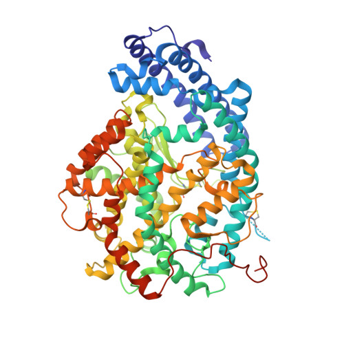
    Angiotensin-I converting enzyme (ACE) is a zinc metalloprotease consisting of two catalytic domains (N- and C-). Most clinical ACE inhibitor(s) (ACEi) have been shown to inhibit both domains nonselectively, resulting in adverse effects such as cough and angioedema. Selectively inhibiting the individual domains is likely to reduce these effects and potentially treat fibrosis in addition to hypertension. ACEi from the GVK Biosciences database were inspected for possible N-domain selective binding patterns. From this set, a diprolyl chemical series was modeled using docking simulations. The series was expanded based on key target interactions involving residues known to impart N-domain selectivity. In total, seven diprolyl compounds were synthesized and tested for N-domain selective ACE inhibition. One compound with an aspartic acid in the P 2 position (compound 16) displayed potent inhibition (K i = 11.45 nM) and was 84-fold more selective toward the N-domain. A high-resolution crystal structure of compound 16 in complex with the N-domain revealed the molecular basis for the observed selectivity.


  • Organizational Affiliation

    Department of Chemistry, University of Cape Town , Rondebosch 7701, South Africa.


Macromolecules
Find similar proteins by:  (by identity cutoff)  |  3D Structure
Entity ID: 1
MoleculeChains Sequence LengthOrganismDetailsImage
Angiotensin-converting enzyme
A, B, C, D
629Homo sapiensMutation(s): 0 
Gene Names: ACEDCPDCP1
EC: 3.2.1 (PDB Primary Data), 3.4.15.1 (PDB Primary Data)
UniProt & NIH Common Fund Data Resources
Find proteins for P12821 (Homo sapiens)
Explore P12821 
Go to UniProtKB:  P12821
PHAROS:  P12821
GTEx:  ENSG00000159640 
Entity Groups  
Sequence Clusters30% Identity50% Identity70% Identity90% Identity95% Identity100% Identity
UniProt GroupP12821
Sequence Annotations
Expand
  • Reference Sequence
Oligosaccharides

Help

Entity ID: 2
MoleculeChains Length2D Diagram Glycosylation3D Interactions
2-acetamido-2-deoxy-beta-D-glucopyranose-(1-4)-2-acetamido-2-deoxy-beta-D-glucopyranose
E, H, M
2N-Glycosylation
Glycosylation Resources
GlyTouCan:  G42666HT
GlyCosmos:  G42666HT
GlyGen:  G42666HT
Entity ID: 3
MoleculeChains Length2D Diagram Glycosylation3D Interactions
beta-D-mannopyranose-(1-4)-2-acetamido-2-deoxy-beta-D-glucopyranose-(1-4)-[alpha-L-fucopyranose-(1-6)]2-acetamido-2-deoxy-beta-D-glucopyranose
F, I, N
4N-Glycosylation
Glycosylation Resources
GlyTouCan:  G32152BH
GlyCosmos:  G32152BH
GlyGen:  G32152BH
Entity ID: 4
MoleculeChains Length2D Diagram Glycosylation3D Interactions
2-acetamido-2-deoxy-beta-D-glucopyranose-(1-4)-[alpha-L-fucopyranose-(1-6)]2-acetamido-2-deoxy-beta-D-glucopyranose
G, L
3N-Glycosylation
Glycosylation Resources
GlyTouCan:  G21290RB
GlyCosmos:  G21290RB
GlyGen:  G21290RB
Entity ID: 5
MoleculeChains Length2D Diagram Glycosylation3D Interactions
alpha-L-fucopyranose-(1-6)-2-acetamido-2-deoxy-beta-D-glucopyranose
J, O
2N-Glycosylation
Glycosylation Resources
GlyTouCan:  G86851RC
GlyCosmos:  G86851RC
GlyGen:  G86851RC
Entity ID: 6
MoleculeChains Length2D Diagram Glycosylation3D Interactions
beta-D-mannopyranose-(1-4)-2-acetamido-2-deoxy-beta-D-glucopyranose-(1-4)-2-acetamido-2-deoxy-beta-D-glucopyranose
K
3N-Glycosylation
Glycosylation Resources
GlyTouCan:  G15407YE
GlyCosmos:  G15407YE
GlyGen:  G15407YE
Small Molecules
Ligands 12 Unique
IDChains Name / Formula / InChI Key2D Diagram3D Interactions
XPE
Query on XPE

Download Ideal Coordinates CCD File 
MB [auth C],
XA [auth B]
3,6,9,12,15,18,21,24,27-NONAOXANONACOSANE-1,29-DIOL
C20 H42 O11
DTPCFIHYWYONMD-UHFFFAOYSA-N
BJ2
Query on BJ2

Download Ideal Coordinates CCD File 
AB [auth C],
CA [auth B],
OB [auth D],
Q [auth A]
(2~{S})-1-[(2~{S})-2-[[(1~{S})-1-[(2~{S})-1-[(2~{S})-2-azanyl-4-oxidanyl-4-oxidanylidene-butanoyl]pyrrolidin-2-yl]-2-oxidanyl-2-oxidanylidene-ethyl]amino]propanoyl]pyrrolidine-2-carboxylic acid
C18 H28 N4 O8
AEFOOLCGQAWEBH-JNLQPACOSA-N
2PE
Query on 2PE

Download Ideal Coordinates CCD File 
WA [auth B]NONAETHYLENE GLYCOL
C18 H38 O10
YZUUTMGDONTGTN-UHFFFAOYSA-N
NAG
Query on NAG

Download Ideal Coordinates CCD File 
YA [auth C]2-acetamido-2-deoxy-beta-D-glucopyranose
C8 H15 N O6
OVRNDRQMDRJTHS-FMDGEEDCSA-N
BCN
Query on BCN

Download Ideal Coordinates CCD File 
AA [auth A]BICINE
C6 H13 N O4
FSVCELGFZIQNCK-UHFFFAOYSA-N
PGE
Query on PGE

Download Ideal Coordinates CCD File 
HC [auth D]
IC [auth D]
JC [auth D]
UA [auth B]
VA [auth B]
HC [auth D],
IC [auth D],
JC [auth D],
UA [auth B],
VA [auth B],
Z [auth A]
TRIETHYLENE GLYCOL
C6 H14 O4
ZIBGPFATKBEMQZ-UHFFFAOYSA-N
PEG
Query on PEG

Download Ideal Coordinates CCD File 
DB [auth C]
EB [auth C]
FA [auth B]
GA [auth B]
HA [auth B]
DB [auth C],
EB [auth C],
FA [auth B],
GA [auth B],
HA [auth B],
IA [auth B],
RB [auth D],
SB [auth D],
T [auth A],
U [auth A]
DI(HYDROXYETHYL)ETHER
C4 H10 O3
MTHSVFCYNBDYFN-UHFFFAOYSA-N
ZN
Query on ZN

Download Ideal Coordinates CCD File 
BA [auth B],
NB [auth D],
P [auth A],
ZA [auth C]
ZINC ION
Zn
PTFCDOFLOPIGGS-UHFFFAOYSA-N
EDO
Query on EDO

Download Ideal Coordinates CCD File 
AC [auth D]
BC [auth D]
FB [auth C]
GB [auth C]
HB [auth C]
AC [auth D],
BC [auth D],
FB [auth C],
GB [auth C],
HB [auth C],
IB [auth C],
JA [auth B],
KA [auth B],
LA [auth B],
MA [auth B],
NA [auth B],
OA [auth B],
PA [auth B],
QA [auth B],
TB [auth D],
UB [auth D],
V [auth A],
VB [auth D],
W [auth A],
WB [auth D],
XB [auth D],
YB [auth D],
ZB [auth D]
1,2-ETHANEDIOL
C2 H6 O2
LYCAIKOWRPUZTN-UHFFFAOYSA-N
ACT
Query on ACT

Download Ideal Coordinates CCD File 
CC [auth D]
DC [auth D]
EC [auth D]
FC [auth D]
GC [auth D]
CC [auth D],
DC [auth D],
EC [auth D],
FC [auth D],
GC [auth D],
JB [auth C],
KB [auth C],
LB [auth C],
RA [auth B],
SA [auth B],
TA [auth B],
X [auth A],
Y [auth A]
ACETATE ION
C2 H3 O2
QTBSBXVTEAMEQO-UHFFFAOYSA-M
CL
Query on CL

Download Ideal Coordinates CCD File 
BB [auth C],
DA [auth B],
PB [auth D],
R [auth A]
CHLORIDE ION
Cl
VEXZGXHMUGYJMC-UHFFFAOYSA-M
MG
Query on MG

Download Ideal Coordinates CCD File 
CB [auth C],
EA [auth B],
QB [auth D],
S [auth A]
MAGNESIUM ION
Mg
JLVVSXFLKOJNIY-UHFFFAOYSA-N
Binding Affinity Annotations 
IDSourceBinding Affinity
BJ2 Binding MOAD:  6EN6 Ki: 11.45 (nM) from 1 assay(s)
Experimental Data & Validation

Experimental Data

  • Method: X-RAY DIFFRACTION
  • Resolution: 1.80 Å
  • R-Value Free: 0.226 
  • R-Value Work: 0.193 
  • R-Value Observed: 0.194 
  • Space Group: P 1
Unit Cell:
Length ( Å )Angle ( ˚ )
a = 74.445α = 84.48
b = 103.373β = 85.5
c = 115.646γ = 81.61
Software Package:
Software NamePurpose
PHENIXrefinement
DIALSdata reduction
Aimlessdata scaling
PHASERphasing

Structure Validation

View Full Validation Report


Ligand Structure Quality Assessment 


Entry History & Funding Information

Deposition Data


Funding OrganizationLocationGrant Number
Medical Research Council (United Kingdom)United KingdomMR/M026647/1

Revision History  (Full details and data files)

  • Version 1.0: 2018-03-07
    Type: Initial release
  • Version 2.0: 2020-07-29
    Type: Remediation
    Reason: Carbohydrate remediation
    Changes: Advisory, Atomic model, Data collection, Derived calculations, Structure summary
  • Version 2.1: 2024-01-17
    Changes: Data collection, Database references, Derived calculations, Refinement description, Structure summary