6EJX | pdb_00006ejx

The metal ion-dependent adhesion site (MIDAS) of the alphaMbeta2 integrin Mac-1 I-domain promiscuously and competitively binds multiple ligands in the regulation of Leukocyte function


Experimental Data Snapshot

  • Method: X-RAY DIFFRACTION
  • Resolution: 2.00 Å
  • R-Value Free: 
    0.232 (Depositor), 0.239 (DCC) 
  • R-Value Work: 
    0.184 (Depositor), 0.196 (DCC) 
  • R-Value Observed: 
    0.185 (Depositor) 

Starting Model: experimental
View more details

wwPDB Validation 3D Report Full Report

Validation slider image for 6EJX

This is version 1.3 of the entry. See complete history

Literature

Structural basis of the leukocyte integrin Mac-1 I-domain interactions with the platelet glycoprotein Ib.

Morgan, J.Saleem, M.Ng, R.Armstrong, C.Wong, S.S.Caulton, S.G.Fickling, A.Williams, H.E.L.Munday, A.D.Lopez, J.A.Searle, M.S.Emsley, J.

(2019) Blood Adv 3: 1450-1459

  • DOI: https://doi.org/10.1182/bloodadvances.2018027011
  • Primary Citation Related Structures: 
    6EJX

  • PubMed Abstract: 

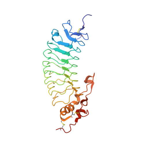
    Cell-surface receptor interactions between leukocyte integrin macrophage-1 antigen (Mac-1, also known as CR3, αMβ2, CD11b/CD18) and platelet glycoprotein Ibα (GPIbα) are critical to vascular inflammation. To define the key residues at the binding interface, we used nuclear magnetic resonance (NMR) to assign the spectra of the mouse Mac-1 I-domain and mapped the residues contacting the mouse GPIbα N-terminal domain (GPIbαN) to the locality of the integrin metal ion-dependant adhesion site (MIDAS) surface. We next determined the crystal structures of the mouse GPIbαN and Mac-1 I-domain to 2 Å and 2.5 Å resolution, respectively. The mouse Mac-1 I-domain crystal structure reveals an active conformation that is stabilized by a crystal contact from the α7-helix with a glutamate side chain completing the octahedral coordination sphere of the MIDAS Mg 2+ ion. The amino acid sequence of the α7-helix and disposition of the glutamic acid matches the C-terminal capping region α-helix of GPIbα effectively acting as a ligand mimetic. Using these crystal structures in combination with NMR measurements and docking analysis, we developed a model whereby an acidic residue from the GPIbα leucine-rich repeat (LRR) capping α-helix coordinates directly to the Mac-1 MIDAS Mg 2+ ion. The Mac-1:GPIbαN complex involves additional interactions consolidated by an elongated pocket flanking the GPIbαN LRR capping α-helix. The GPIbαN α-helix has an HxxxE motif, which is equivalent by homology to RxxxD from the human GPIbαN. Subsequent mutagenesis of residues at this interface, coupled with surface plasmon resonance studies, confirmed the importance of GPIbαN residues H218, E222, and the Mac-1 MIDAS residue T209 to formation of the complex.


  • Organizational Affiliation
    • School of Chemistry and.

Macromolecule Content 

  • Total Structure Weight: 63.55 kDa 
  • Atom Count: 4,803 
  • Modeled Residue Count: 532 
  • Deposited Residue Count: 556 
  • Unique protein chains: 1

Macromolecules

Find similar proteins by:|  3D Structure
Entity ID: 1
MoleculeChains  Sequence LengthOrganismDetailsImage
Platelet glycoprotein Ib alpha chainA [auth D],
B [auth A]
278Mus musculusMutation(s): 0 
Gene Names: Gp1ba
UniProt
Find proteins for O35930 (Mus musculus)
Explore O35930 
Go to UniProtKB:  O35930
Entity Groups
Sequence Clusters30% Identity50% Identity70% Identity90% Identity95% Identity100% Identity
UniProt GroupO35930
Sequence Annotations
Expand
Reference Sequence

Small Molecules

Ligands 5 Unique
IDChains Name / Formula / InChI Key2D Diagram3D Interactions
MES

Query on MES


Download:Ideal Coordinates CCD File
E [auth D],
O [auth A]
2-(N-MORPHOLINO)-ETHANESULFONIC ACID
C6 H13 N O4 S
SXGZJKUKBWWHRA-UHFFFAOYSA-N
PEG

Query on PEG


Download:Ideal Coordinates CCD File
D,
G [auth A],
H [auth A]
DI(HYDROXYETHYL)ETHER
C4 H10 O3
MTHSVFCYNBDYFN-UHFFFAOYSA-N
GOL

Query on GOL


Download:Ideal Coordinates CCD File
I [auth A],
J [auth A]
GLYCEROL
C3 H8 O3
PEDCQBHIVMGVHV-UHFFFAOYSA-N
IMD

Query on IMD


Download:Ideal Coordinates CCD File
C [auth D],
F [auth A]
IMIDAZOLE
C3 H5 N2
RAXXELZNTBOGNW-UHFFFAOYSA-O
POL

Query on POL


Download:Ideal Coordinates CCD File
K [auth A],
L [auth A],
M [auth A],
N [auth A]
N-PROPANOL
C3 H8 O
BDERNNFJNOPAEC-UHFFFAOYSA-N

Experimental Data & Validation

Experimental Data

  • Method: X-RAY DIFFRACTION
  • Resolution: 2.00 Å
  • R-Value Free:  0.232 (Depositor), 0.239 (DCC) 
  • R-Value Work:  0.184 (Depositor), 0.196 (DCC) 
  • R-Value Observed: 0.185 (Depositor) 
Space Group: P 21 21 21
Unit Cell:
Length ( Å )Angle ( ˚ )
a = 61.537α = 90
b = 72.87β = 90
c = 164.053γ = 90
Software Package:
Software NamePurpose
REFMACrefinement
xia2data reduction
Aimlessdata scaling
MOLREPmodel building

Structure Validation

View Full Validation Report


Entry History 

Deposition Data

Revision History  (Full details and data files)

  • Version 1.0: 2018-10-24
    Type: Initial release
  • Version 1.1: 2019-11-06
    Changes: Data collection, Database references
  • Version 1.2: 2024-01-17
    Changes: Advisory, Data collection, Database references, Refinement description
  • Version 1.3: 2024-11-13
    Changes: Structure summary