Self-Assembling 2D Arrays with de Novo Protein Building Blocks.
Chen, Z., Johnson, M.C., Chen, J., Bick, M.J., Boyken, S.E., Lin, B., De Yoreo, J.J., Kollman, J.M., Baker, D., DiMaio, F.(2019) J Am Chem Soc 141: 8891-8895
- PubMed: 31050411 Search on PubMedSearch on PubMed Central
- DOI: https://doi.org/10.1021/jacs.9b01978
- Primary Citation Related Structures:
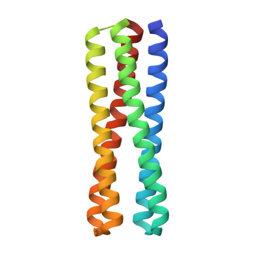
6EGC - PubMed Abstract:
Modular self-assembly of biomolecules in two dimensions (2D) is straightforward with DNA but has been difficult to realize with proteins, due to the lack of modular specificity similar to Watson-Crick base pairing. Here we describe a general approach to design 2D arrays using de novo designed pseudosymmetric protein building blocks. A homodimeric helical bundle was reconnected into a monomeric building block, and the surface was redesigned in Rosetta to enable self-assembly into a 2D array in the C12 layer symmetry group. Two out of ten designed arrays assembled to micrometer scale under negative stain electron microscopy, and displayed the designed lattice geometry with assembly size up to 100 nm under atomic force microscopy. The design of 2D arrays with pseudosymmetric building blocks is an important step toward the design of programmable protein self-assembly via pseudosymmetric patterning of orthogonal binding interfaces.
- Physical Sciences Division , Pacific Northwest National Laboratory , Richland , Washington 99352 , United States.
Organizational Affiliation: