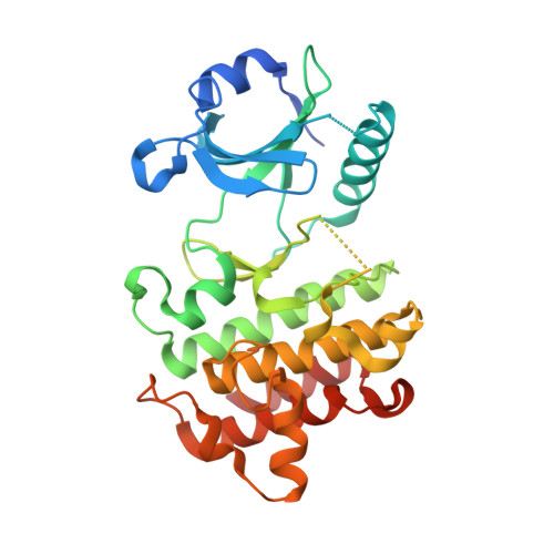
6EG9

IRAK4 in complex with Ponatinib


Experimental Data Snapshot

  • Method: X-RAY DIFFRACTION
  • Resolution: 2.41 Å
  • R-Value Free: 0.249 
  • R-Value Work: 0.223 
  • R-Value Observed: 0.224 

wwPDB Validation   3D Report Full Report


Ligand Structure Quality Assessment 


This is version 1.3 of the entry. See complete history


Literature

Conformational flexibility and inhibitor binding to unphosphorylated interleukin-1 receptor-associated kinase 4 (IRAK4).

Wang, L.Ferrao, R.Li, Q.Hatcher, J.M.Choi, H.G.Buhrlage, S.J.Gray, N.S.Wu, H.

(2019) J Biol Chem 294: 4511-4519

  • DOI: https://doi.org/10.1074/jbc.RA118.005428
  • Primary Citation of Related Structures:  
    6EG9, 6EGA, 6EGD, 6EGE, 6EGF

  • PubMed Abstract: 

    Interleukin-1 receptor-associated kinase 4 (IRAK4) is a key player in innate immune and inflammatory responses, performing a critical role in signal transduction downstream of Toll-like receptors and interleukin-1 (IL-1) receptors. Upon ligand binding and via its N-terminal death domain, IRAK4 is recruited to an oligomeric receptor that is proximal to the Myddosome signaling complex, inducing IRAK4 kinase domain dimerization, autophosphorylation, and activation. To date, all known IRAK4 structures are in the active conformation, precluding a good understanding of IRAK4's conformational dynamics. To address this issue, here we first solved three crystal structures of the IRAK4 kinase domain (at ≤2.6 Å resolution), in its unphosphorylated, inactive state bound to either the ATP analog AMP-PNP or to one of the two small-molecule inhibitors JH-I-25 and JH-I-17. The structures disclosed that although the structure in complex with AMP-PNP is in an "αC-out" inactive conformation, those in complex with type I inhibitors assume an active "Asp-Phe-Gly (DFG)-in" and "αC-in" conformation. The ability of unphosphorylated IRAK4 to take on variable conformations prompted us to screen for small-molecule inhibitors that bind preferentially to unphosphorylated IRAK4, leading to the identification of ponatinib and HG-12-6. Solving the structures of unphosphorylated IRAK4 in complex with these two inhibitors, we found that they both bind as type II inhibitors with IRAK4 in a "DFG-out" conformation. Collectively, these structures reveal conformational flexibility of unphosphorylated IRAK4 and provide unexpected insights into the potential use of small molecules to modulate IRAK4 activity in cancer, autoimmunity, and inflammation.


  • Organizational Affiliation

    From the Department of Biological Chemistry and Molecular Pharmacology, Harvard Medical School, Boston, Massachusetts 02115.


Macromolecules
Find similar proteins by:  (by identity cutoff)  |  3D Structure
Entity ID: 1
MoleculeChains Sequence LengthOrganismDetailsImage
Interleukin-1 receptor-associated kinase 4
A, B
312Homo sapiensMutation(s): 0 
Gene Names: IRAK4
EC: 2.7.11.1
UniProt & NIH Common Fund Data Resources
Find proteins for Q9NWZ3 (Homo sapiens)
Explore Q9NWZ3 
Go to UniProtKB:  Q9NWZ3
PHAROS:  Q9NWZ3
GTEx:  ENSG00000198001 
Entity Groups  
Sequence Clusters30% Identity50% Identity70% Identity90% Identity95% Identity100% Identity
UniProt GroupQ9NWZ3
Sequence Annotations
Expand
  • Reference Sequence
Small Molecules
Ligands 3 Unique
IDChains Name / Formula / InChI Key2D Diagram3D Interactions
0LI (Subject of Investigation/LOI)
Query on 0LI

Download Ideal Coordinates CCD File 
C [auth A],
F [auth B]
3-(imidazo[1,2-b]pyridazin-3-ylethynyl)-4-methyl-N-{4-[(4-methylpiperazin-1-yl)methyl]-3-(trifluoromethyl)phenyl}benzam ide
C29 H27 F3 N6 O
PHXJVRSECIGDHY-UHFFFAOYSA-N
SO4
Query on SO4

Download Ideal Coordinates CCD File 
D [auth A],
G [auth B]
SULFATE ION
O4 S
QAOWNCQODCNURD-UHFFFAOYSA-L
CO
Query on CO

Download Ideal Coordinates CCD File 
E [auth A],
H [auth B],
I [auth B],
J [auth B]
COBALT (II) ION
Co
XLJKHNWPARRRJB-UHFFFAOYSA-N
Experimental Data & Validation

Experimental Data

  • Method: X-RAY DIFFRACTION
  • Resolution: 2.41 Å
  • R-Value Free: 0.249 
  • R-Value Work: 0.223 
  • R-Value Observed: 0.224 
  • Space Group: P 61 2 2
Unit Cell:
Length ( Å )Angle ( ˚ )
a = 85.037α = 90
b = 85.037β = 90
c = 434.301γ = 120
Software Package:
Software NamePurpose
PHENIXrefinement
XDSdata reduction
XDSdata scaling
PHASERphasing

Structure Validation

View Full Validation Report


Ligand Structure Quality Assessment 


Entry History & Funding Information

Deposition Data


Funding OrganizationLocationGrant Number
National Institutes of Health/National Institute Of Allergy and Infectious Diseases (NIH/NIAID)United StatesAI050872

Revision History  (Full details and data files)

  • Version 1.0: 2019-02-20
    Type: Initial release
  • Version 1.1: 2019-09-04
    Changes: Data collection, Database references
  • Version 1.2: 2019-12-18
    Changes: Author supporting evidence
  • Version 1.3: 2024-03-13
    Changes: Data collection, Database references, Derived calculations, Structure summary