


Crystal Structure of Ribose-5-phosphate Isomerase from Legionella pneumophila
Braverman, K.N., Dranow, D.M., Mayclin, S.J., Lorimer, D.D., Horanyi, P.S., Edwards, T.E.To be published.
Experimental Data Snapshot
Starting Model: experimental
View more details
wwPDB Validation 3D Report Full Report

Entity ID: 1 | |||||
|---|---|---|---|---|---|
| Molecule | Chains | Sequence Length | Organism | Details | Image |
| Ribose-5-phosphate isomerase A | 218 | Legionella pneumophila subsp. pneumophila str. Philadelphia 1 | Mutation(s): 0 Gene Names: rpiA, lpg0094 EC: 5.3.1.6 | 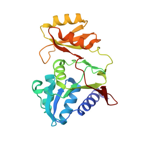 | |
UniProt | |||||
Entity Groups | |||||
| Sequence Clusters | 30% Identity50% Identity70% Identity90% Identity95% Identity100% Identity | ||||
| UniProt Group | Q5ZZB7 | ||||
Sequence AnnotationsExpand | |||||
Reference Sequence | |||||
| Ligands 1 Unique | |||||
|---|---|---|---|---|---|
| ID | Chains | Name / Formula / InChI Key | 2D Diagram | 3D Interactions | |
| NA Download:Ideal Coordinates CCD File | C [auth A], D [auth B] | SODIUM ION Na FKNQFGJONOIPTF-UHFFFAOYSA-N |  | ||
| Length ( Å ) | Angle ( ˚ ) |
|---|---|
| a = 104.63 | α = 90 |
| b = 67.93 | β = 119.54 |
| c = 74 | γ = 90 |
| Software Name | Purpose |
|---|---|
| PHENIX | refinement |
| XDS | data reduction |
| XSCALE | data scaling |
| PDB_EXTRACT | data extraction |
| MoRDa | phasing |


