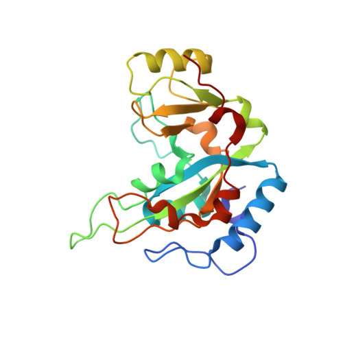
6EDG

Pseudomonas exotoxin A domain III T18H477L


Experimental Data Snapshot

  • Method: X-RAY DIFFRACTION
  • Resolution: 1.47 Å
  • R-Value Free: 0.192 
  • R-Value Work: 0.177 
  • R-Value Observed: 0.178 

wwPDB Validation   3D Report Full Report


Ligand Structure Quality Assessment 


This is version 1.3 of the entry. See complete history


Literature

Deimmunizing substitutions in Pseudomonasexotoxin domain III perturb antigen processing without eliminating T-cell epitopes.

Moss, D.L.Park, H.W.Mettu, R.R.Landry, S.J.

(2019) J Biol Chem 294: 4667-4681

  • DOI: https://doi.org/10.1074/jbc.RA118.006704
  • Primary Citation of Related Structures:  
    6EDG

  • PubMed Abstract: 

    Effective adaptive immune responses depend on activation of CD4+ T cells via the presentation of antigen peptides in the context of major histocompatibility complex (MHC) class II. The structure of an antigen strongly influences its processing within the endolysosome and potentially controls the identity of peptides that are presented to T cells. A recombinant immunotoxin, comprising exotoxin A domain III (PE-III) from Pseudomonas aeruginosa and a cancer-specific antibody fragment, has been developed to manage cancer, but its effectiveness is limited by the induction of neutralizing antibodies. Here, we observed that this immunogenicity is substantially reduced by substituting six residues within PE-III. Although these substitutions targeted T-cell epitopes, we demonstrate that reduced conformational stability and protease resistance were responsible for the reduced antibody titer. Analysis of mouse T-cell responses coupled with biophysical studies on single-substitution versions of PE-III suggested that modest but comprehensible changes in T-cell priming can dramatically perturb antibody production. The most strongly responsive PE-III epitope was well-predicted by a structure-based algorithm. In summary, single-residue substitutions can drastically alter the processing and immunogenicity of PE-III but have only modest effects on CD4+ T-cell priming in mice. Our findings highlight the importance of structure-based processing constraints for accurate epitope prediction.


  • Organizational Affiliation

    From the Department of Biochemistry and Molecular Biology, Tulane University School of Medicine, New Orleans, Louisiana 70112 and.


Macromolecules
Find similar proteins by:  (by identity cutoff)  |  3D Structure
Entity ID: 1
MoleculeChains Sequence LengthOrganismDetailsImage
Exotoxin213Pseudomonas aeruginosaMutation(s): 5 
Gene Names: CGU42_27545
Entity Groups  
Sequence Clusters30% Identity50% Identity70% Identity90% Identity95% Identity100% Identity
Sequence Annotations
Expand
  • Reference Sequence
Small Molecules
Ligands 1 Unique
IDChains Name / Formula / InChI Key2D Diagram3D Interactions
P34
Query on P34

Download Ideal Coordinates CCD File 
B [auth A]N~2~,N~2~-DIMETHYL-N~1~-(6-OXO-5,6-DIHYDROPHENANTHRIDIN-2-YL)GLYCINAMIDE
C17 H17 N3 O2
UYJZZVDLGDDTCL-UHFFFAOYSA-N
Experimental Data & Validation

Experimental Data

  • Method: X-RAY DIFFRACTION
  • Resolution: 1.47 Å
  • R-Value Free: 0.192 
  • R-Value Work: 0.177 
  • R-Value Observed: 0.178 
  • Space Group: P 21 21 21
Unit Cell:
Length ( Å )Angle ( ˚ )
a = 38.373α = 90
b = 48.188β = 90
c = 99.002γ = 90
Software Package:
Software NamePurpose
REFMACrefinement
XDSdata reduction
SCALAdata scaling
MOLREPphasing

Structure Validation

View Full Validation Report


Ligand Structure Quality Assessment 


Entry History & Funding Information

Deposition Data


Funding OrganizationLocationGrant Number
National Institutes of Health/National Institute Of Allergy and Infectious Diseases (NIH/NIAID)United StatesR21-AI122199

Revision History  (Full details and data files)

  • Version 1.0: 2019-02-06
    Type: Initial release
  • Version 1.1: 2019-08-21
    Changes: Data collection, Database references
  • Version 1.2: 2019-12-18
    Changes: Author supporting evidence
  • Version 1.3: 2023-10-11
    Changes: Data collection, Database references, Refinement description