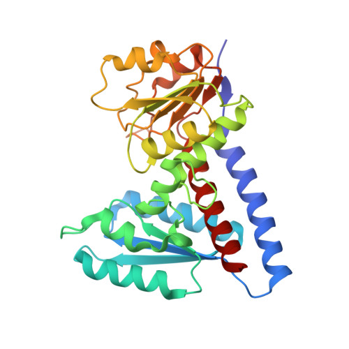
6ECR

The human methylenetetrahydrofolate dehydrogenase/cyclohydrolase (FolD) complexed with NADP


Experimental Data Snapshot

  • Method: X-RAY DIFFRACTION
  • Resolution: 2.20 Å
  • R-Value Free: 0.219 
  • R-Value Work: 0.170 
  • R-Value Observed: 0.175 

wwPDB Validation   3D Report Full Report


Ligand Structure Quality Assessment 


This is version 1.2 of the entry. See complete history

Re-refinement Note

This entry reflects an alternative modeling of the original data in: 1DIG


Literature

An assessment of three human methylenetetrahydrofolate dehydrogenase/cyclohydrolase-ligand complexes following further refinement.

Bueno, R.Dawson, A.Hunter, W.N.

(2019) Acta Crystallogr F Struct Biol Commun 75: 148-152

  • DOI: https://doi.org/10.1107/S2053230X18018083
  • Primary Citation of Related Structures:  
    6ECP, 6ECQ, 6ECR

  • PubMed Abstract: 

    The enzymes involved in folate metabolism are key drug targets for cell-growth modulation, and accurate crystallographic structures provide templates to be exploited for structure-based ligand design. In this context, three ternary complex structures of human methylenetetrahydrofolate dehydrogenase/cyclohydrolase have been published [Schmidt et al. (2000), Biochemistry, 39, 6325-6335] and potentially represent starting points for the development of new antifolate inhibitors. However, an inspection of the models and the deposited data revealed deficiencies and raised questions about the validity of the structures. A number of inconsistencies relating to the publication were also identified. Additional refinement was carried out with the deposited data, seeking to improve the models and to then validate the complex structures or correct the record. In one case, the inclusion of the inhibitor in the structure was supported and alterations to the model allowed details of enzyme-ligand interactions to be described that had not previously been discussed. For one weak inhibitor, the data suggested that the ligand may adopt two poses in the binding site, both with few interactions with the enzyme. In the third case, that of a potent inhibitor, inconsistencies were noted in the assignment of the chemical structure and there was no evidence to support the inclusion of the ligand in the active site.


  • Organizational Affiliation

    Division of Biological Chemistry and Drug Discovery, College of Life Sciences, University of Dundee, Dundee DD1 5EH, Scotland.


Macromolecules
Find similar proteins by:  (by identity cutoff)  |  3D Structure
Entity ID: 1
MoleculeChains Sequence LengthOrganismDetailsImage
methylenetetrahydrofolate dehydrogenase cyclohydrolase
A, B
296Homo sapiensMutation(s): 0 
Gene Names: MTHFD1MTHFCMTHFD
EC: 1.5.1.5 (PDB Primary Data), 3.5.4.9 (PDB Primary Data), 6.3.4.3 (PDB Primary Data)
UniProt & NIH Common Fund Data Resources
Find proteins for P11586 (Homo sapiens)
Explore P11586 
Go to UniProtKB:  P11586
PHAROS:  P11586
GTEx:  ENSG00000100714 
Entity Groups  
Sequence Clusters30% Identity50% Identity70% Identity90% Identity95% Identity100% Identity
UniProt GroupP11586
Sequence Annotations
Expand
  • Reference Sequence
Experimental Data & Validation

Experimental Data

  • Method: X-RAY DIFFRACTION
  • Resolution: 2.20 Å
  • R-Value Free: 0.219 
  • R-Value Work: 0.170 
  • R-Value Observed: 0.175 
  • Space Group: P 21 21 21
Unit Cell:
Length ( Å )Angle ( ˚ )
a = 67.889α = 90
b = 136.419β = 90
c = 61.579γ = 90
Software Package:
Software NamePurpose
REFMACrefinement
DENZOdata reduction
SCALEPACKdata scaling
REFMACphasing

Structure Validation

View Full Validation Report


Ligand Structure Quality Assessment 


Entry History & Funding Information

Deposition Data


Funding OrganizationLocationGrant Number
Sao Paulo Research Foundation (FAPESP)Brazil2016/16038-5

Revision History  (Full details and data files)

  • Version 1.0: 2019-03-27
    Type: Initial release
  • Version 1.1: 2020-01-01
    Changes: Author supporting evidence
  • Version 1.2: 2023-10-11
    Changes: Data collection, Database references, Refinement description