


Crystal structure of Leishmania major dihydroorotate dehydrogenase mutant H174A in complex with orotate
Reis, R.A.G., Pinheiro, M.P., de Souza, A.L., Hunter, W.N., Nonato, M.C.To be published.
Experimental Data Snapshot
Starting Model: experimental
View more details
Entity ID: 1 | |||||
|---|---|---|---|---|---|
| Molecule | Chains | Sequence Length | Organism | Details | Image |
| Dihydroorotate dehydrogenase (fumarate) | 354 | Leishmania major | Mutation(s): 1 Gene Names: DHODH, LMJF_16_0530 EC: 1.3.98.1 | 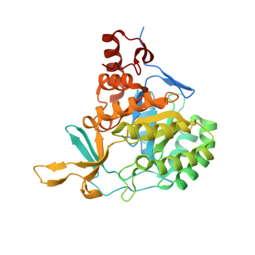 | |
UniProt | |||||
Entity Groups | |||||
| Sequence Clusters | 30% Identity50% Identity70% Identity90% Identity95% Identity100% Identity | ||||
| UniProt Group | Q4QEW7 | ||||
Sequence AnnotationsExpand | |||||
Reference Sequence | |||||
| Ligands 5 Unique | |||||
|---|---|---|---|---|---|
| ID | Chains | Name / Formula / InChI Key | 2D Diagram | 3D Interactions | |
| FMN Download:Ideal Coordinates CCD File | C [auth A], K [auth B] | FLAVIN MONONUCLEOTIDE C17 H21 N4 O9 P FVTCRASFADXXNN-SCRDCRAPSA-N |  | ||
| ORO Download:Ideal Coordinates CCD File | D [auth A], L [auth B] | OROTIC ACID C5 H4 N2 O4 PXQPEWDEAKTCGB-UHFFFAOYSA-N |  | ||
| SO4 Download:Ideal Coordinates CCD File | H [auth A] I [auth A] J [auth A] N [auth B] O [auth B] | SULFATE ION O4 S QAOWNCQODCNURD-UHFFFAOYSA-L |  | ||
| GOL Download:Ideal Coordinates CCD File | E [auth A], F [auth A], G [auth A], M [auth B] | GLYCEROL C3 H8 O3 PEDCQBHIVMGVHV-UHFFFAOYSA-N |  | ||
| NI Download:Ideal Coordinates CCD File | Q [auth B] | NICKEL (II) ION Ni VEQPNABPJHWNSG-UHFFFAOYSA-N |  | ||
| Length ( Å ) | Angle ( ˚ ) |
|---|---|
| a = 144.279 | α = 90 |
| b = 144.279 | β = 90 |
| c = 69.913 | γ = 120 |
| Software Name | Purpose |
|---|---|
| Aimless | data scaling |
| MOLREP | phasing |
| REFMAC | refinement |
| PDB_EXTRACT | data extraction |
| Funding Organization | Location | Grant Number |
|---|---|---|
| Sao Paulo Research Foundation (FAPESP) | Brazil | 11/23504-9 |
| Sao Paulo Research Foundation (FAPESP) | Brazil | 07/08703-0 |


