


Structural Basis for Respiratory Syncytial Virus Fusion Inhibitor Resistance
Battles, M.B., McLellan, J.S.To be published.
Experimental Data Snapshot
Starting Model: experimental
View more details
Entity ID: 1 | |||||
|---|---|---|---|---|---|
| Molecule | Chains | Sequence Length | Organism | Details | Image |
| Fusion glycoprotein F0 | A [auth F] | 568 | human respiratory syncytial virus | Mutation(s): 5 | 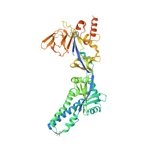 |
UniProt | |||||
Entity Groups | |||||
| Sequence Clusters | 30% Identity50% Identity70% Identity90% Identity95% Identity100% Identity | ||||
| UniProt Group | P03420 | ||||
Glycosylation | |||||
| Glycosylation Sites: 1 | |||||
Sequence AnnotationsExpand | |||||
Reference Sequence | |||||
| Ligands 3 Unique | |||||
|---|---|---|---|---|---|
| ID | Chains | Name / Formula / InChI Key | 2D Diagram | 3D Interactions | |
| NAG Download:Ideal Coordinates CCD File | B [auth F] | 2-acetamido-2-deoxy-beta-D-glucopyranose C8 H15 N O6 OVRNDRQMDRJTHS-FMDGEEDCSA-N |  | ||
| NHE Download:Ideal Coordinates CCD File | C [auth F], D [auth F] | 2-[N-CYCLOHEXYLAMINO]ETHANE SULFONIC ACID C8 H17 N O3 S MKWKNSIESPFAQN-UHFFFAOYSA-N |  | ||
| SO4 Download:Ideal Coordinates CCD File | E [auth F], F, G [auth F], H [auth F], I [auth F] | SULFATE ION O4 S QAOWNCQODCNURD-UHFFFAOYSA-L |  | ||
| Length ( Å ) | Angle ( ˚ ) |
|---|---|
| a = 168.73 | α = 90 |
| b = 168.73 | β = 90 |
| c = 168.73 | γ = 90 |
| Software Name | Purpose |
|---|---|
| Aimless | data scaling |
| PHENIX | refinement |
| PDB_EXTRACT | data extraction |
| MOSFLM | data reduction |
| PHASER | phasing |
| Funding Organization | Location | Grant Number |
|---|---|---|
| National Institutes of Health/National Institute Of Allergy and Infectious Diseases (NIH/NIAID) | United States | 5T32AI007519-18 |
| National Institutes of Health/National Institute of General Medical Sciences (NIH/NIGMS) | United States | P20GM113132 |


