6E7Z | pdb_00006e7z

cryo-EM structure of human TRPML1 with ML-SA1 and PI35P2


Experimental Data Snapshot

  • Method: ELECTRON MICROSCOPY
  • Resolution: 3.73 Å
  • Aggregation State: PARTICLE 
  • Reconstruction Method: SINGLE PARTICLE 

wwPDB Validation 3D Report Full Report

Validation slider image for 6E7Z

This is version 1.2 of the entry. See complete history

Literature

Structural basis for PtdInsP2-mediated human TRPML1 regulation.

Fine, M.Schmiege, P.Li, X.

(2018) Nat Commun 9: 4192-4192

  • DOI: https://doi.org/10.1038/s41467-018-06493-7
  • Primary Citation Related Structures: 
    6E7P, 6E7Y, 6E7Z

  • PubMed Abstract: 

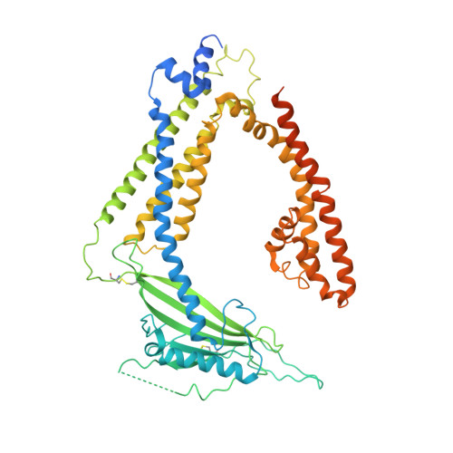
    Transient receptor potential mucolipin 1 (TRPML1), a lysosomal channel, maintains the low pH and calcium levels for lysosomal function. Several small molecules modulate TRPML1 activity. ML-SA1, a synthetic agonist, binds to the pore region and phosphatidylinositol-3,5-bisphosphate (PtdIns(3,5)P 2 ), a natural lipid, stimulates channel activity to a lesser extent than ML-SA1; moreover, PtdIns(4,5)P 2 , another natural lipid, prevents TRPML1-mediated calcium release. Notably, PtdIns(3,5)P 2 and ML-SA1 cooperate further increasing calcium efflux. Here we report the structures of human TRPML1 at pH 5.0 with PtdIns(3,5)P 2 , PtdIns(4,5)P 2 , or ML-SA1 and PtdIns(3,5)P 2 , revealing a unique lipid-binding site. PtdIns(3,5)P 2 and PtdIns(4,5)P 2 bind to the extended helices of S1, S2, and S3. The phosphate group of PtdIns(3,5)P 2 induces Y355 to form a π-cation interaction with R403, moving the S4-S5 linker, thus allosterically activating the channel. Our structures and electrophysiological characterizations reveal an allosteric site and provide molecular insight into how lipids regulate TRP channels.


  • Organizational Affiliation
    • Department of Physiology, University of Texas Southwestern Medical Center, Dallas, TX, 75390, USA.

Macromolecule Content 

  • Total Structure Weight: 264.79 kDa 
  • Atom Count: 15,616 
  • Modeled Residue Count: 1,900 
  • Deposited Residue Count: 2,320 
  • Unique protein chains: 1

Macromolecules

Find similar proteins by:|  3D Structure
Entity ID: 1
MoleculeChains  Sequence LengthOrganismDetailsImage
Mucolipin-1
A, B, C, D
580Homo sapiensMutation(s): 0 
Gene Names: MCOLN1ML4MSTP080
Membrane Entity: Yes 
UniProt & NIH Common Fund Data Resources
Find proteins for Q9GZU1 (Homo sapiens)
Explore Q9GZU1 
Go to UniProtKB:  Q9GZU1
PHAROS:  Q9GZU1
GTEx:  ENSG00000090674 
Entity Groups
Sequence Clusters30% Identity50% Identity70% Identity90% Identity95% Identity100% Identity
UniProt GroupQ9GZU1
Sequence Annotations
Expand
Reference Sequence

Small Molecules

Ligands 2 Unique
IDChains Name / Formula / InChI Key2D Diagram3D Interactions
HZ7

Query on HZ7


Download:Ideal Coordinates CCD File
E [auth A],
G [auth B],
I [auth C],
K [auth D]
(1R,2S,3S,4R,5S,6R)-5-{[(R)-[(2R)-2,3-bis{[(1S)-1-hydroxyoctyl]oxy}propoxy](hydroxy)phosphoryl]oxy}-2,4,6-trihydroxycyclohexane-1,3-diyl bis[dihydrogen (phosphate)]
C25 H53 O19 P3
YMIVTWICVQDVIN-OZVNAFTMSA-N
AQV

Query on AQV


Download:Ideal Coordinates CCD File
F [auth A],
H [auth B],
J [auth C],
L [auth D]
2-{2-oxo-2-[(4S)-2,2,4-trimethyl-3,4-dihydroquinolin-1(2H)-yl]ethyl}-1H-isoindole-1,3(2H)-dione
C22 H22 N2 O3
KDDHBJICVBONAX-AWEZNQCLSA-N

Experimental Data & Validation

Experimental Data

  • Method: ELECTRON MICROSCOPY
  • Resolution: 3.73 Å
  • Aggregation State: PARTICLE 
  • Reconstruction Method: SINGLE PARTICLE 
EM Software:
TaskSoftware PackageVersion
MODEL REFINEMENTPHENIX
RECONSTRUCTIONFREALIX

Structure Validation

View Full Validation Report


Entry History 

Deposition Data

Revision History  (Full details and data files)

  • Version 1.0: 2018-11-28
    Type: Initial release
  • Version 1.1: 2024-11-20
    Changes: Data collection, Database references, Structure summary
  • Version 1.2: 2025-05-21
    Changes: Data collection