6E7O

Crystal structure of deglycosylated human EPDR1

  • Classification: LIPID BINDING PROTEIN
  • Organism(s): Homo sapiens
  • Expression System: Homo sapiens
  • Mutation(s): No 

  • Deposited: 2018-07-27 Released: 2019-01-23 
  • Deposition Author(s): Wei, Y., Prive, G.G.
  • Funding Organization(s): Canadian Institutes of Health Research (CIHR), Natural Sciences and Engineering Research Council (NSERC, Canada)

Experimental Data Snapshot

  • Method: X-RAY DIFFRACTION
  • Resolution: 3.00 Å
  • R-Value Free: 0.278 
  • R-Value Work: 0.255 
  • R-Value Observed: 0.256 

wwPDB Validation   3D Report Full Report


This is version 1.2 of the entry. See complete history


Literature

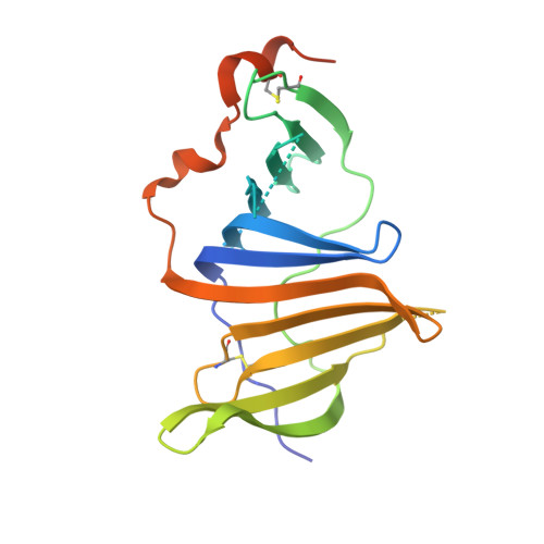
Crystal structures of human lysosomal EPDR1 reveal homology with the superfamily of bacterial lipoprotein transporters.

Wei, Y.Xiong, Z.J.Li, J.Zou, C.Cairo, C.W.Klassen, J.S.Prive, G.G.

(2019) Commun Biol 2: 52-52

  • DOI: https://doi.org/10.1038/s42003-018-0262-9
  • Primary Citation of Related Structures:  
    6E7O, 6E8N

  • PubMed Abstract: 

    EPDR1, a member of the ependymin-related protein family, is a relatively uncharacterized protein found in the lysosomes and secretomes of most vertebrates. Despite having roles in human disease and health, the molecular functions of EPDR1 remain unknown. Here, we present crystal structures of human EPDR1 and reveal that the protein adopts a fold previously seen only in bacterial proteins related to the LolA lipoprotein transporter. EPDR1 forms a homodimer with an overall shape resembling a half-shell with two non-overlapping hydrophobic grooves on the flat side of the hemisphere. EPDR1 can interact with membranes that contain negatively charged lipids, including BMP and GM1, and we suggest that EPDR1 may function as a lysosomal activator protein or a lipid transporter. A phylogenetic analysis reveals that the fold is more widely distributed than previously suspected, with representatives identified in all branches of cellular life.


  • Organizational Affiliation

    1Princess Margaret Cancer Centre, Toronto, M5G 1L7 ON Canada.


Macromolecules
Find similar proteins by:  (by identity cutoff)  |  3D Structure
Entity ID: 1
MoleculeChains Sequence LengthOrganismDetailsImage
Mammalian ependymin-related protein 1
A, B, C, D
194Homo sapiensMutation(s): 0 
Gene Names: EPDR1MERP1UCC1
UniProt & NIH Common Fund Data Resources
Find proteins for Q9UM22 (Homo sapiens)
Explore Q9UM22 
Go to UniProtKB:  Q9UM22
PHAROS:  Q9UM22
GTEx:  ENSG00000086289 
Entity Groups  
Sequence Clusters30% Identity50% Identity70% Identity90% Identity95% Identity100% Identity
UniProt GroupQ9UM22
Sequence Annotations
Expand
  • Reference Sequence
Experimental Data & Validation

Experimental Data

  • Method: X-RAY DIFFRACTION
  • Resolution: 3.00 Å
  • R-Value Free: 0.278 
  • R-Value Work: 0.255 
  • R-Value Observed: 0.256 
  • Space Group: C 2 2 21
Unit Cell:
Length ( Å )Angle ( ˚ )
a = 87.805α = 90
b = 97.482β = 90
c = 189.469γ = 90
Software Package:
Software NamePurpose
PHENIXrefinement
HKL-2000data reduction
Aimlessdata scaling
PHENIXphasing

Structure Validation

View Full Validation Report


Entry History & Funding Information

Deposition Data


Funding OrganizationLocationGrant Number
Canadian Institutes of Health Research (CIHR)Canada--
Natural Sciences and Engineering Research Council (NSERC, Canada)Canada--

Revision History  (Full details and data files)

  • Version 1.0: 2019-01-23
    Type: Initial release
  • Version 1.1: 2019-02-20
    Changes: Data collection, Database references
  • Version 1.2: 2020-01-08
    Changes: Author supporting evidence