6E7D | pdb_00006e7d

Structure of the inhibitory NKR-P1B receptor bound to the host-encoded ligand, Clr-b


Experimental Data Snapshot

  • Method: X-RAY DIFFRACTION
  • Resolution: 2.90 Å
  • R-Value Free: 
    0.225 (Depositor), 0.223 (DCC) 
  • R-Value Work: 
    0.186 (Depositor), 0.205 (DCC) 
  • R-Value Observed: 
    0.188 (Depositor) 

Starting Models: experimental
View more details

wwPDB Validation 3D Report Full Report

Validation slider image for 6E7D

Ligand Structure Quality Assessment 


This is version 1.5 of the entry. See complete history

Literature

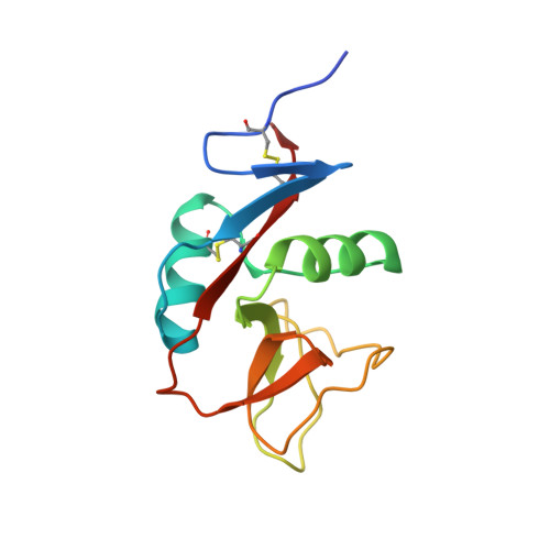
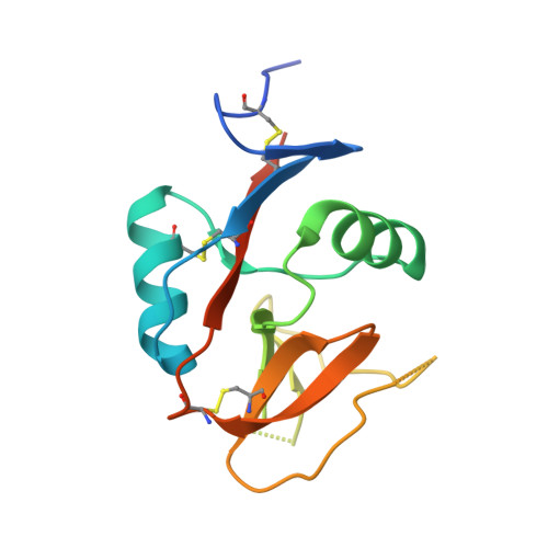
Recognition of host Clr-b by the inhibitory NKR-P1B receptor provides a basis for missing-self recognition.

Balaji, G.R.Aguilar, O.A.Tanaka, M.Shingu-Vazquez, M.A.Fu, Z.Gully, B.S.Lanier, L.L.Carlyle, J.R.Rossjohn, J.Berry, R.

(2018) Nat Commun 9: 4623-4623

  • DOI: https://doi.org/10.1038/s41467-018-06989-2
  • Primary Citation Related Structures: 
    6E7D

  • PubMed Abstract: 

    The interaction between natural killer (NK) cell inhibitory receptors and their cognate ligands constitutes a key mechanism by which healthy tissues are protected from NK cell-mediated lysis. However, self-ligand recognition remains poorly understood within the prototypical NKR-P1 receptor family. Here we report the structure of the inhibitory NKR-P1B receptor bound to its cognate host ligand, Clr-b. NKR-P1B and Clr-b interact via a head-to-head docking mode through an interface that includes a large array of polar interactions. NKR-P1B:Clr-b recognition is extremely sensitive to mutations at the heterodimeric interface, with most mutations severely impacting both Clr-b binding and NKR-P1B receptor function to implicate a low affinity interaction. Within the structure, two NKR-P1B:Clr-b complexes are cross-linked by a non-classic NKR-P1B homodimer, and the disruption of homodimer formation abrogates Clr-b recognition. These data provide an insight into a fundamental missing-self recognition system and suggest an avidity-based mechanism underpins NKR-P1B receptor function.


  • Organizational Affiliation
    • Infection and Immunity Program and Department of Biochemistry and Molecular Biology, Biomedicine Discovery Institute, Monash University, Clayton, Victoria, 3800, Australia.

Macromolecule Content 

  • Total Structure Weight: 363.9 kDa 
  • Atom Count: 22,989 
  • Modeled Residue Count: 2,877 
  • Deposited Residue Count: 3,080 
  • Unique protein chains: 2

Macromolecules

Find similar proteins by:|  3D Structure
Entity ID: 1
MoleculeChains  Sequence LengthOrganismDetailsImage
C-type lectin domain family 2 member D
A, B, C, D, E
A, B, C, D, E, F, G, H, I, J, K, L, M, N, O, P
124Mus musculusMutation(s): 0 
Gene Names: Clec2dClrbOcil
UniProt
Find proteins for Q91V08 (Mus musculus)
Explore Q91V08 
Go to UniProtKB:  Q91V08
Entity Groups
Sequence Clusters30% Identity50% Identity70% Identity90% Identity95% Identity100% Identity
UniProt GroupQ91V08
Sequence Annotations
Expand
Reference Sequence
Find similar proteins by:|  3D Structure
Entity ID: 2
MoleculeChains  Sequence LengthOrganismDetailsImage
Killer cell lectin-like receptor subfamily B member 1B allele B
Q, R, S, T, U
Q, R, S, T, U, V, W, X
137Mus musculusMutation(s): 1 
Gene Names: Klrb1bKlrb1dLy55dNkrp1bNkrp1d
UniProt
Find proteins for Q99JB4 (Mus musculus)
Explore Q99JB4 
Go to UniProtKB:  Q99JB4
Entity Groups
Sequence Clusters30% Identity50% Identity70% Identity90% Identity95% Identity100% Identity
UniProt GroupQ99JB4
Glycosylation
Glycosylation Sites: 1
Sequence Annotations
Expand
Reference Sequence

Small Molecules

Ligands 2 Unique
IDChains Name / Formula / InChI Key2D Diagram3D Interactions
NAG

Query on NAG


Download:Ideal Coordinates CCD File
BA [auth R]
DA [auth S]
FA [auth T]
HA [auth U]
JA [auth V]
BA [auth R],
DA [auth S],
FA [auth T],
HA [auth U],
JA [auth V],
LA [auth W],
NA [auth X],
Z [auth Q]
2-acetamido-2-deoxy-beta-D-glucopyranose
C8 H15 N O6
OVRNDRQMDRJTHS-FMDGEEDCSA-N
SO4

Query on SO4


Download:Ideal Coordinates CCD File
AA [auth Q]
CA [auth R]
EA [auth S]
GA [auth T]
IA [auth U]
AA [auth Q],
CA [auth R],
EA [auth S],
GA [auth T],
IA [auth U],
KA [auth V],
MA [auth W],
OA [auth X],
Y [auth D]
SULFATE ION
O4 S
QAOWNCQODCNURD-UHFFFAOYSA-L

Experimental Data & Validation

Experimental Data

  • Method: X-RAY DIFFRACTION
  • Resolution: 2.90 Å
  • R-Value Free:  0.225 (Depositor), 0.223 (DCC) 
  • R-Value Work:  0.186 (Depositor), 0.205 (DCC) 
  • R-Value Observed: 0.188 (Depositor) 
Space Group: P 1
Unit Cell:
Length ( Å )Angle ( ˚ )
a = 66.93α = 73.12
b = 122.069β = 82.08
c = 131.561γ = 84.46
Software Package:
Software NamePurpose
BUSTERrefinement
iMOSFLMdata reduction
SCALAdata scaling
MOLREPphasing

Structure Validation

View Full Validation Report


Ligand Structure Quality Assessment 


Entry History 

& Funding Information

Deposition Data


Funding OrganizationLocationGrant Number
National Health and Medical Research Council (NHMRC, Australia)AustraliaAPP1109901
Australian Research Council (ARC)AustraliaFL160100049

Revision History  (Full details and data files)

  • Version 1.0: 2018-10-24
    Type: Initial release
  • Version 1.1: 2019-05-08
    Changes: Data collection, Database references
  • Version 1.2: 2020-01-01
    Changes: Author supporting evidence
  • Version 1.3: 2020-07-29
    Type: Remediation
    Reason: Carbohydrate remediation
    Changes: Data collection, Derived calculations, Structure summary
  • Version 1.4: 2023-10-11
    Changes: Data collection, Database references, Refinement description, Structure summary
  • Version 1.5: 2024-10-30
    Changes: Structure summary