6DVS | pdb_00006dvs

Crystal structure of Pseudomonas stutzeri D-phenylglycine aminotransferase


Experimental Data Snapshot

  • Method: X-RAY DIFFRACTION
  • Resolution: 1.82 Å
  • R-Value Free: 
    0.222 (Depositor), 0.224 (DCC) 
  • R-Value Work: 
    0.179 (Depositor), 0.180 (DCC) 
  • R-Value Observed: 
    0.181 (Depositor) 

wwPDB Validation 3D Report Full Report

Validation slider image for 6DVS

This is version 1.2 of the entry. See complete history

Literature

Structural Determinants of the Stereoinverting Activity of Pseudomonas stutzeri d-Phenylglycine Aminotransferase.

Walton, C.J.W.Thiebaut, F.Brunzelle, J.S.Couture, J.F.Chica, R.A.

(2018) Biochemistry 57: 5437-5446

  • DOI: https://doi.org/10.1021/acs.biochem.8b00767
  • Primary Citation Related Structures: 
    6DVS

  • PubMed Abstract: 

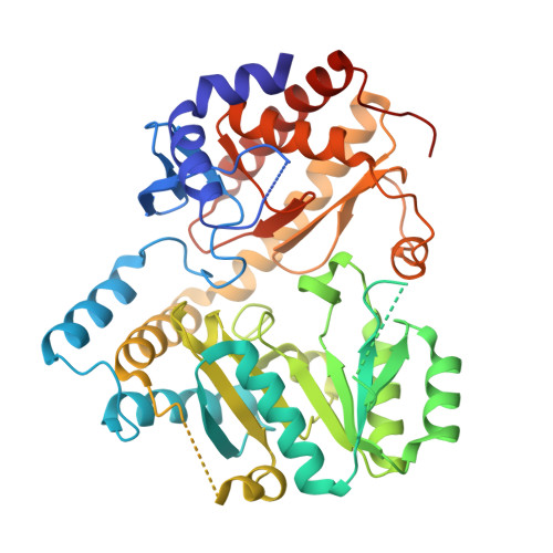
    Aromatic d-amino acids are key precursors for the production of many small molecule therapeutics. Therefore, the development of biocatalytic methods for their synthesis is of great interest. An enzyme that has great potential as a biocatalyst for the synthesis of d-amino acids is the stereoinverting d-phenylglycine aminotransferase (DPAT) from Pseudomonas stutzeri ST-201. This enzyme catalyzes a unique l to d transamination reaction that produces d-phenylglycine and α-ketoglutarate from benzoylformate and l-glutamate, via a mechanism that is poorly understood. Here, we present the crystal structure of DPAT, which shows that the enzyme folds into a two-domain structure representative of class III aminotransferases. Guided by the crystal structure, we performed saturation mutagenesis to probe the substrate binding pockets of the enzyme. These experiments helped us identify two arginine residues (R34 and R407), one in each binding pocket, that are essential to catalysis. Together with kinetic analyses using a library of amino acid substrates, our mutagenesis and structural studies allow us to propose a binding model that explains the dual l/d specificity of DPAT. Our kinetic analyses also demonstrate that DPAT can catalyze the transamination of β- and γ-amino acids, reclassifying this enzyme as an ω-aminotransferase. Collectively, our studies highlight that the DPAT active site is amenable to protein engineering for expansion of its substrate scope, which offers the opportunity to generate new biocatalysts for the synthesis of a variety of valuable optically pure d-amino acids from inexpensive and abundant l-amino acids.


  • Organizational Affiliation
    • Department of Chemistry and Biomolecular Sciences , University of Ottawa , Ottawa , Ontario , Canada K1N 6N5.

Macromolecule Content 

  • Total Structure Weight: 50.64 kDa 
  • Atom Count: 3,591 
  • Modeled Residue Count: 397 
  • Deposited Residue Count: 453 
  • Unique protein chains: 1

Macromolecules

Find similar proteins by:|  3D Structure
Entity ID: 1
MoleculeChains  Sequence LengthOrganismDetailsImage
D-phenylglycine aminotransferase453Stutzerimonas stutzeriMutation(s): 0 
Gene Names: dpgA
EC: 2.6.1.72
UniProt
Find proteins for Q6VY99 (Stutzerimonas stutzeri)
Explore Q6VY99 
Go to UniProtKB:  Q6VY99
Entity Groups
Sequence Clusters30% Identity50% Identity70% Identity90% Identity95% Identity100% Identity
UniProt GroupQ6VY99
Sequence Annotations
Expand
Reference Sequence

Small Molecules

Ligands 5 Unique
IDChains Name / Formula / InChI Key2D Diagram3D Interactions
TRS

Query on TRS


Download:Ideal Coordinates CCD File
S [auth A]2-AMINO-2-HYDROXYMETHYL-PROPANE-1,3-DIOL
C4 H12 N O3
LENZDBCJOHFCAS-UHFFFAOYSA-O
SO4

Query on SO4


Download:Ideal Coordinates CCD File
B [auth A]
C [auth A]
D [auth A]
E [auth A]
F [auth A]
B [auth A],
C [auth A],
D [auth A],
E [auth A],
F [auth A],
G [auth A]
SULFATE ION
O4 S
QAOWNCQODCNURD-UHFFFAOYSA-L
PO4

Query on PO4


Download:Ideal Coordinates CCD File
T [auth A]PHOSPHATE ION
O4 P
NBIIXXVUZAFLBC-UHFFFAOYSA-K
EDO

Query on EDO


Download:Ideal Coordinates CCD File
H [auth A]
I [auth A]
J [auth A]
K [auth A]
L [auth A]
H [auth A],
I [auth A],
J [auth A],
K [auth A],
L [auth A],
M [auth A],
N [auth A],
O [auth A],
P [auth A],
Q [auth A],
R [auth A]
1,2-ETHANEDIOL
C2 H6 O2
LYCAIKOWRPUZTN-UHFFFAOYSA-N
ACT

Query on ACT


Download:Ideal Coordinates CCD File
U [auth A]ACETATE ION
C2 H3 O2
QTBSBXVTEAMEQO-UHFFFAOYSA-M

Experimental Data & Validation

Experimental Data

  • Method: X-RAY DIFFRACTION
  • Resolution: 1.82 Å
  • R-Value Free:  0.222 (Depositor), 0.224 (DCC) 
  • R-Value Work:  0.179 (Depositor), 0.180 (DCC) 
  • R-Value Observed: 0.181 (Depositor) 
Space Group: P 31 2 1
Unit Cell:
Length ( Å )Angle ( ˚ )
a = 74.639α = 90
b = 74.639β = 90
c = 149.948γ = 120
Software Package:
Software NamePurpose
PHENIXrefinement
XDSdata reduction
Aimlessdata scaling
PHASERphasing

Structure Validation

View Full Validation Report


Entry History 

Deposition Data

Revision History  (Full details and data files)

  • Version 1.0: 2018-09-12
    Type: Initial release
  • Version 1.1: 2018-10-03
    Changes: Data collection, Database references
  • Version 1.2: 2024-03-13
    Changes: Advisory, Data collection, Database references