6DV3 | pdb_00006dv3

Structure of the Salmonella SPI-1 type III secretion injectisome secretin InvG in the open gate state


Experimental Data Snapshot

  • Method: ELECTRON MICROSCOPY
  • Resolution: 4.10 Å
  • Aggregation State: PARTICLE 
  • Reconstruction Method: SINGLE PARTICLE 

wwPDB Validation 3D Report Full Report

Validation slider image for 6DV3

This is version 1.2 of the entry. See complete history

Literature

Cryo-EM analysis of the T3S injectisome reveals the structure of the needle and open secretin.

Hu, J.Worrall, L.J.Hong, C.Vuckovic, M.Atkinson, C.E.Caveney, N.Yu, Z.Strynadka, N.C.J.

(2018) Nat Commun 9: 3840-3840

  • DOI: https://doi.org/10.1038/s41467-018-06298-8
  • Primary Citation Related Structures: 
    6DUZ, 6DV3, 6DV6, 6DWB

  • PubMed Abstract: 

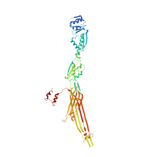
    The bacterial type III secretion system, or injectisome, is a syringe shaped nanomachine essential for the virulence of many disease causing Gram-negative bacteria. At the core of the injectisome structure is the needle complex, a continuous channel formed by the highly oligomerized inner and outer membrane hollow rings and a polymerized helical needle filament which spans through and projects into the infected host cell. Here we present the near-atomic resolution structure of a needle complex from the prototypical Salmonella Typhimurium SPI-1 type III secretion system, with local masking protocols allowing for model building and refinement of the major membrane spanning components of the needle complex base in addition to an isolated needle filament. This work provides significant insight into injectisome structure and assembly and importantly captures the molecular basis for substrate induced gating in the giant outer membrane secretin portal family.


  • Organizational Affiliation
    • Department of Biochemistry and Molecular Biology and the Centre for Blood Research, University of British Columbia, Vancouver, V6T 1Z3, BC, Canada.

Macromolecule Content 

  • Total Structure Weight: 927.53 kDa 
  • Atom Count: 57,975 
  • Modeled Residue Count: 7,500 
  • Deposited Residue Count: 8,430 
  • Unique protein chains: 1

Macromolecules

Find similar proteins by:|  3D Structure
Entity ID: 1
MoleculeChains  Sequence LengthOrganismDetailsImage
Protein InvG
A, B, C, D, E
A, B, C, D, E, F, G, H, I, J, K, L, M, N, O
562Salmonella enterica subsp. enterica serovar TyphimuriumMutation(s): 0 
Membrane Entity: Yes 
UniProt
Find proteins for P35672 (Salmonella typhimurium (strain LT2 / SGSC1412 / ATCC 700720))
Explore P35672 
Go to UniProtKB:  P35672
Entity Groups
Sequence Clusters30% Identity50% Identity70% Identity90% Identity95% Identity100% Identity
UniProt GroupP35672
Sequence Annotations
Expand
Reference Sequence

Experimental Data & Validation

Experimental Data

  • Method: ELECTRON MICROSCOPY
  • Resolution: 4.10 Å
  • Aggregation State: PARTICLE 
  • Reconstruction Method: SINGLE PARTICLE 
EM Software:
TaskSoftware PackageVersion
MODEL REFINEMENTPHENIX1.13
RECONSTRUCTIONRELION2.01

Structure Validation

View Full Validation Report


Entry History 

& Funding Information

Deposition Data


Funding OrganizationLocationGrant Number
Canadian Institutes of Health Research (CIHR)Canada--
Howard Hughes Medical Institute (HHMI)United States--

Revision History  (Full details and data files)

  • Version 1.0: 2018-10-03
    Type: Initial release
  • Version 1.1: 2019-11-20
    Changes: Author supporting evidence
  • Version 1.2: 2024-03-13
    Changes: Data collection, Database references