Force-dependent allostery of the alpha-catenin actin-binding domain controls adherens junction dynamics and functions.
Ishiyama, N., Sarpal, R., Wood, M.N., Barrick, S.K., Nishikawa, T., Hayashi, H., Kobb, A.B., Flozak, A.S., Yemelyanov, A., Fernandez-Gonzalez, R., Yonemura, S., Leckband, D.E., Gottardi, C.J., Tepass, U., Ikura, M.(2018) Nat Commun 9: 5121-5121
- PubMed: 30504777 Search on PubMedSearch on PubMed Central
- DOI: https://doi.org/10.1038/s41467-018-07481-7
- Primary Citation Related Structures:
6DUW, 6DUY, 6DV1 - PubMed Abstract:
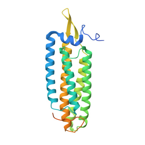
α-catenin is a key mechanosensor that forms force-dependent interactions with F-actin, thereby coupling the cadherin-catenin complex to the actin cytoskeleton at adherens junctions (AJs). However, the molecular mechanisms by which α-catenin engages F-actin under tension remained elusive. Here we show that the α1-helix of the α-catenin actin-binding domain (αcat-ABD) is a mechanosensing motif that regulates tension-dependent F-actin binding and bundling. αcat-ABD containing an α1-helix-unfolding mutation (H1) shows enhanced binding to F-actin in vitro. Although full-length α-catenin-H1 can generate epithelial monolayers that resist mechanical disruption, it fails to support normal AJ regulation in vivo. Structural and simulation analyses suggest that α1-helix allosterically controls the actin-binding residue V796 dynamics. Crystal structures of αcat-ABD-H1 homodimer suggest that α-catenin can facilitate actin bundling while it remains bound to E-cadherin. We propose that force-dependent allosteric regulation of αcat-ABD promotes dynamic interactions with F-actin involved in actin bundling, cadherin clustering, and AJ remodeling during tissue morphogenesis.
- Princess Margaret Cancer Centre, University Health Network, Toronto, ON, M5G 1L7, Canada. noboru.ishiyama@uhnresearch.ca.
Organizational Affiliation: