6DS1 | pdb_00006ds1

Crystal structure of Cj0485 dehydrogenase in complex with NADP+


Experimental Data Snapshot

  • Method: X-RAY DIFFRACTION
  • Resolution: 2.12 Å
  • R-Value Free: 
    0.214 (Depositor), 0.213 (DCC) 
  • R-Value Work: 
    0.159 (Depositor), 0.159 (DCC) 
  • R-Value Observed: 
    0.162 (Depositor) 

Starting Model: experimental
View more details

wwPDB Validation 3D Report Full Report

Validation slider image for 6DS1

Ligand Structure Quality Assessment 


This is version 1.2 of the entry. See complete history

Literature

The gastrointestinal pathogen Campylobacter jejuni metabolizes sugars with potential help from commensal Bacteroides vulgatus.

Garber, J.M.Nothaft, H.Pluvinage, B.Stahl, M.Bian, X.Porfirio, S.Enriquez, A.Butcher, J.Huang, H.Glushka, J.Line, E.Gerlt, J.A.Azadi, P.Stintzi, A.Boraston, A.B.Szymanski, C.M.

(2020) Commun Biol 3: 2-2

  • DOI: https://doi.org/10.1038/s42003-019-0727-5
  • Primary Citation Related Structures: 
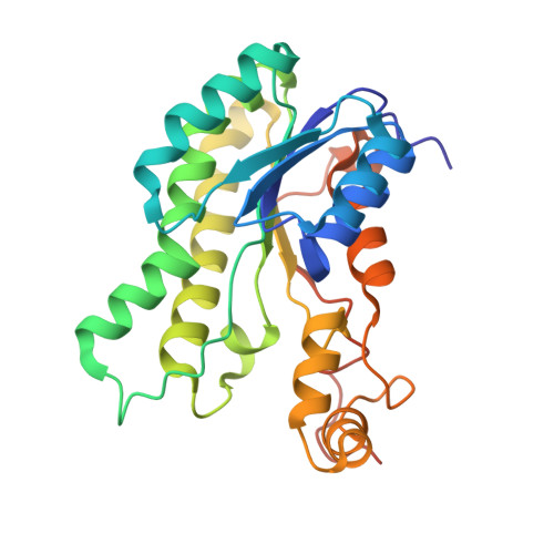
    6DRR, 6DS1

  • PubMed Abstract: 

    Although the gastrointestinal pathogen Campylobacter jejuni was considered asaccharolytic, >50% of sequenced isolates possess an operon for L-fucose utilization. In C. jejuni NCTC11168, this pathway confers L-fucose chemotaxis and competitive colonization advantages in the piglet diarrhea model, but the catabolic steps remain unknown. Here we solved the putative dehydrogenase structure, resembling FabG of Burkholderia multivorans. The C. jejuni enzyme, FucX, reduces L-fucose and D-arabinose in vitro and both sugars are catabolized by fuc-operon encoded enzymes. This enzyme alone confers chemotaxis to both sugars in a non-carbohydrate-utilizing C. jejuni strain. Although C. jejuni lacks fucosidases, the organism exhibits enhanced growth in vitro when co-cultured with Bacteroides vulgatus, suggesting scavenging may occur. Yet, when excess amino acids are available, C. jejuni prefers them to carbohydrates, indicating a metabolic hierarchy exists. Overall this study increases understanding of nutrient metabolism by this pathogen, and identifies interactions with other gut microbes.


  • Organizational Affiliation
    • Department of Microbiology, University of Georgia, 527 Biological Sciences Bldg, Athens, GA, 30602, USA.

Macromolecule Content 

  • Total Structure Weight: 125.43 kDa 
  • Atom Count: 8,798 
  • Modeled Residue Count: 1,037 
  • Deposited Residue Count: 1,084 
  • Unique protein chains: 1

Macromolecules

Find similar proteins by:|  3D Structure
Entity ID: 1
MoleculeChains  Sequence LengthOrganismDetailsImage
Putative oxidoreductase
A, B, C, D
271Campylobacter jejuni subsp. jejuni NCTC 11168 = ATCC 700819Mutation(s): 0 
Gene Names: Cj0485
UniProt
Find proteins for Q0PB28 (Campylobacter jejuni subsp. jejuni serotype O:2 (strain ATCC 700819 / NCTC 11168))
Explore Q0PB28 
Go to UniProtKB:  Q0PB28
Entity Groups
Sequence Clusters30% Identity50% Identity70% Identity90% Identity95% Identity100% Identity
UniProt GroupQ0PB28
Sequence Annotations
Expand
Reference Sequence

Small Molecules

Ligands 3 Unique
IDChains Name / Formula / InChI Key2D Diagram3D Interactions
NAP

Query on NAP


Download:Ideal Coordinates CCD File
E [auth A],
H [auth B],
J [auth C],
L [auth D]
NADP NICOTINAMIDE-ADENINE-DINUCLEOTIDE PHOSPHATE
C21 H28 N7 O17 P3
XJLXINKUBYWONI-NNYOXOHSSA-N
GOL

Query on GOL


Download:Ideal Coordinates CCD File
F [auth A],
M [auth D],
N [auth D]
GLYCEROL
C3 H8 O3
PEDCQBHIVMGVHV-UHFFFAOYSA-N
MG

Query on MG


Download:Ideal Coordinates CCD File
G [auth A],
I [auth B],
K [auth C],
O [auth D]
MAGNESIUM ION
Mg
JLVVSXFLKOJNIY-UHFFFAOYSA-N

Experimental Data & Validation

Experimental Data

  • Method: X-RAY DIFFRACTION
  • Resolution: 2.12 Å
  • R-Value Free:  0.214 (Depositor), 0.213 (DCC) 
  • R-Value Work:  0.159 (Depositor), 0.159 (DCC) 
  • R-Value Observed: 0.162 (Depositor) 
Space Group: P 1
Unit Cell:
Length ( Å )Angle ( ˚ )
a = 68.504α = 89.31
b = 68.462β = 113.43
c = 70.718γ = 115.67
Software Package:
Software NamePurpose
HKL-2000data reduction
HKL-2000data scaling
PHASERphasing
PHENIXrefinement
PDB_EXTRACTdata extraction

Structure Validation

View Full Validation Report


Ligand Structure Quality Assessment 


Entry History 

Deposition Data

Revision History  (Full details and data files)

  • Version 1.0: 2019-07-10
    Type: Initial release
  • Version 1.1: 2020-07-29
    Changes: Database references
  • Version 1.2: 2023-10-11
    Changes: Data collection, Database references, Refinement description