6DRK | pdb_00006drk

Structure of TRPM2 ion channel receptor by single particle electron cryo-microscopy, Apo state


Experimental Data Snapshot

  • Method: ELECTRON MICROSCOPY
  • Resolution: 3.80 Å
  • Aggregation State: PARTICLE 
  • Reconstruction Method: SINGLE PARTICLE 

wwPDB Validation 3D Report Full Report

Validation slider image for 6DRK

This is version 1.4 of the entry. See complete history

Literature

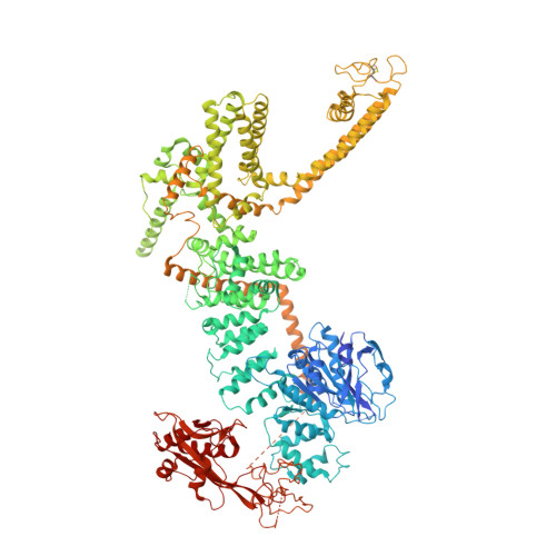
Architecture of the TRPM2 channel and its activation mechanism by ADP-ribose and calcium.

Huang, Y.Winkler, P.A.Sun, W.Lu, W.Du, J.

(2018) Nature 562: 145-149

  • DOI: https://doi.org/10.1038/s41586-018-0558-4
  • Primary Citation Related Structures: 
    6DRJ, 6DRK

  • PubMed Abstract: 

    Transient receptor potential melastatin 2 (TRPM2) is a calcium-permeable, non-selective cation channel that has an essential role in diverse physiological processes such as core body temperature regulation, immune response and apoptosis 1-4 . TRPM2 is polymodal and can be activated by a wide range of stimuli 1-7 , including temperature, oxidative stress and NAD + -related metabolites such as ADP-ribose (ADPR). Its activation results in both Ca 2+ entry across the plasma membrane and Ca 2+ release from lysosomes 8 , and has been linked to diseases such as ischaemia-reperfusion injury, bipolar disorder and Alzheimer's disease 9-11 . Here we report the cryo-electron microscopy structures of the zebrafish TRPM2 in the apo resting (closed) state and in the ADPR/Ca 2+ -bound active (open) state, in which the characteristic NUDT9-H domains hang underneath the MHR1/2 domain. We identify an ADPR-binding site located in the bi-lobed structure of the MHR1/2 domain. Our results provide an insight into the mechanism of activation of the TRPM channel family and define a framework for the development of therapeutic agents to treat neurodegenerative diseases and temperature-related pathological conditions.


  • Organizational Affiliation
    • Van Andel Research Institute, Grand Rapids, MI, USA.

Macromolecule Content 

  • Total Structure Weight: 674.92 kDa 
  • Atom Count: 33,120 
  • Modeled Residue Count: 5,060 
  • Deposited Residue Count: 5,896 
  • Unique protein chains: 1

Macromolecules

Find similar proteins by:|  3D Structure
Entity ID: 1
MoleculeChains  Sequence LengthOrganismDetailsImage
Transient receptor potential cation channel, subfamily M, member 2A,
B [auth C],
C [auth B],
D
1,474Danio rerioMutation(s): 0 
Gene Names: trpm2
Membrane Entity: Yes 
UniProt
Find proteins for A0A0R4IMY7 (Danio rerio)
Explore A0A0R4IMY7 
Go to UniProtKB:  A0A0R4IMY7
Entity Groups
Sequence Clusters30% Identity50% Identity70% Identity90% Identity95% Identity100% Identity
UniProt GroupA0A0R4IMY7
Sequence Annotations
Expand
Reference Sequence

Experimental Data & Validation

Experimental Data

  • Method: ELECTRON MICROSCOPY
  • Resolution: 3.80 Å
  • Aggregation State: PARTICLE 
  • Reconstruction Method: SINGLE PARTICLE 
EM Software:
TaskSoftware PackageVersion
MODEL REFINEMENTPHENIX

Structure Validation

View Full Validation Report


Entry History 

Deposition Data

Revision History  (Full details and data files)

  • Version 1.0: 2018-09-19
    Type: Initial release
  • Version 1.1: 2018-09-26
    Changes: Data collection, Structure summary
  • Version 1.2: 2018-10-10
    Changes: Data collection, Database references, Structure summary
  • Version 1.3: 2024-10-23
    Changes: Data collection, Database references, Structure summary
  • Version 1.4: 2025-05-14
    Changes: Data collection