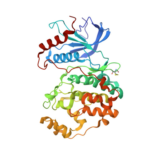
6DMG

A multiconformer ligand model of EK6 bound to ERK2


Experimental Data Snapshot

  • Method: X-RAY DIFFRACTION
  • Resolution: 2.20 Å
  • R-Value Free: 0.236 
  • R-Value Work: 0.186 
  • R-Value Observed: 0.189 

wwPDB Validation   3D Report Full Report


Ligand Structure Quality Assessment 


This is version 1.1 of the entry. See complete history

Re-refinement Note

This entry reflects an alternative modeling of the original data in: 4FV3


Literature

qFit-ligand Reveals Widespread Conformational Heterogeneity of Drug-Like Molecules in X-Ray Electron Density Maps.

van Zundert, G.C.P.Hudson, B.M.de Oliveira, S.H.P.Keedy, D.A.Fonseca, R.Heliou, A.Suresh, P.Borrelli, K.Day, T.Fraser, J.S.van den Bedem, H.

(2018) J Med Chem 61: 11183-11198

  • DOI: https://doi.org/10.1021/acs.jmedchem.8b01292
  • Primary Citation of Related Structures:  
    6DMG, 6DMH, 6DMI, 6DMJ, 6DMK, 6DML

  • PubMed Abstract: 

    Proteins and ligands sample a conformational ensemble that governs molecular recognition, activity, and dissociation. In structure-based drug design, access to this conformational ensemble is critical to understand the balance between entropy and enthalpy in lead optimization. However, ligand conformational heterogeneity is currently severely underreported in crystal structures in the Protein Data Bank, owing in part to a lack of automated and unbiased procedures to model an ensemble of protein-ligand states into X-ray data. Here, we designed a computational method, qFit-ligand, to automatically resolve conformationally averaged ligand heterogeneity in crystal structures, and applied it to a large set of protein receptor-ligand complexes. In an analysis of the cancer related BRD4 domain, we found that up to 29% of protein crystal structures bound with drug-like molecules present evidence of unmodeled, averaged, relatively isoenergetic conformations in ligand-receptor interactions. In many retrospective cases, these alternate conformations were adventitiously exploited to guide compound design, resulting in improved potency or selectivity. Combining qFit-ligand with high-throughput screening or multitemperature crystallography could therefore augment the structure-based drug design toolbox.


  • Organizational Affiliation

    Schrödinger , New York , New York 10036 , United States.


Macromolecules
Find similar proteins by:  (by identity cutoff)  |  3D Structure
Entity ID: 1
MoleculeChains Sequence LengthOrganismDetailsImage
Mitogen-activated protein kinase 1347Homo sapiensMutation(s): 0 
Gene Names: MAPK1ERK2PRKM1PRKM2
EC: 2.7.11.24
UniProt & NIH Common Fund Data Resources
Find proteins for P28482 (Homo sapiens)
Explore P28482 
Go to UniProtKB:  P28482
PHAROS:  P28482
GTEx:  ENSG00000100030 
Entity Groups  
Sequence Clusters30% Identity50% Identity70% Identity90% Identity95% Identity100% Identity
UniProt GroupP28482
Sequence Annotations
Expand
  • Reference Sequence
Small Molecules
Ligands 3 Unique
IDChains Name / Formula / InChI Key2D Diagram3D Interactions
EK6 (Subject of Investigation/LOI)
Query on EK6

Download Ideal Coordinates CCD File 
H [auth A]ethyl N-{2-chloro-4-[5-(5-{[(1S)-1-(3-chloro-4-fluorophenyl)-2-hydroxyethyl]carbamoyl}-1H-pyrrol-3-yl)-1H-pyrazol-4-yl]benzyl}glycinate
C27 H26 Cl2 F N5 O4
VVRLXUNPTACSJC-XMMPIXPASA-N
SO4
Query on SO4

Download Ideal Coordinates CCD File 
B [auth A],
C [auth A],
D [auth A]
SULFATE ION
O4 S
QAOWNCQODCNURD-UHFFFAOYSA-L
EDO
Query on EDO

Download Ideal Coordinates CCD File 
E [auth A],
F [auth A],
G [auth A]
1,2-ETHANEDIOL
C2 H6 O2
LYCAIKOWRPUZTN-UHFFFAOYSA-N
Modified Residues  1 Unique
IDChains TypeFormula2D DiagramParent
CME
Query on CME
A
L-PEPTIDE LINKINGC5 H11 N O3 S2CYS
Experimental Data & Validation

Experimental Data

  • Method: X-RAY DIFFRACTION
  • Resolution: 2.20 Å
  • R-Value Free: 0.236 
  • R-Value Work: 0.186 
  • R-Value Observed: 0.189 
  • Space Group: P 1 21 1
Unit Cell:
Length ( Å )Angle ( ˚ )
a = 48.78α = 90
b = 70.7β = 110.11
c = 60.86γ = 90
Software Package:
Software NamePurpose
PHENIXrefinement
PDB_EXTRACTdata extraction
MOSFLMdata reduction
SCALEPACKdata scaling
PHENIXphasing

Structure Validation

View Full Validation Report


Ligand Structure Quality Assessment 


Entry History 

Deposition Data

Revision History  (Full details and data files)

  • Version 1.0: 2018-12-19
    Type: Initial release
  • Version 1.1: 2019-04-24
    Changes: Data collection, Database references