6DKK

Structure of BoNT


Experimental Data Snapshot

  • Method: X-RAY DIFFRACTION
  • Resolution: 2.70 Å
  • R-Value Free: 0.223 
  • R-Value Work: 0.209 
  • R-Value Observed: 0.210 

wwPDB Validation   3D Report Full Report


This is version 1.2 of the entry. See complete history


Literature

A viral-fusion-peptide-like molecular switch drives membrane insertion of botulinum neurotoxin A1.

Lam, K.H.Guo, Z.Krez, N.Matsui, T.Perry, K.Weisemann, J.Rummel, A.Bowen, M.E.Jin, R.

(2018) Nat Commun 9: 5367-5367

  • DOI: https://doi.org/10.1038/s41467-018-07789-4
  • Primary Citation of Related Structures:  
    6DKK, 6MHJ

  • PubMed Abstract: 

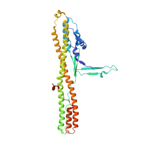
    Botulinum neurotoxin (BoNT) delivers its protease domain across the vesicle membrane to enter the neuronal cytosol upon vesicle acidification. This process is mediated by its translocation domain (H N ), but the molecular mechanism underlying membrane insertion of H N remains poorly understood. Here, we report two crystal structures of BoNT/A1 H N that reveal a novel molecular switch (termed BoNT-switch) in H N , where buried α-helices transform into surface-exposed hydrophobic β-hairpins triggered by acidic pH. Locking the BoNT-switch by disulfide trapping inhibited the association of H N with anionic liposomes, blocked channel formation by H N , and reduced the neurotoxicity of BoNT/A1 by up to ~180-fold. Single particle counting studies showed that an acidic environment tends to promote BoNT/A1 self-association on liposomes, which is partly regulated by the BoNT-switch. These findings suggest that the BoNT-switch flips out upon exposure to the acidic endosomal pH, which enables membrane insertion of H N that subsequently leads to LC delivery.


  • Organizational Affiliation

    Department of Physiology and Biophysics, University of California, Irvine, 92697, CA, USA.


Macromolecules
Find similar proteins by:  (by identity cutoff)  |  3D Structure
Entity ID: 1
MoleculeChains Sequence LengthOrganismDetailsImage
Botulinum neurotoxin type A
A, B
327Clostridium botulinum A str. ATCC 19397Mutation(s): 0 
Gene Names: botAatxbna
EC: 3.4.24.69
UniProt
Find proteins for P0DPI0 (Clostridium botulinum)
Explore P0DPI0 
Go to UniProtKB:  P0DPI0
Entity Groups  
Sequence Clusters30% Identity50% Identity70% Identity90% Identity95% Identity100% Identity
UniProt GroupP0DPI0
Sequence Annotations
Expand
  • Reference Sequence
Small Molecules
Ligands 1 Unique
IDChains Name / Formula / InChI Key2D Diagram3D Interactions
PO4
Query on PO4

Download Ideal Coordinates CCD File 
C [auth B]PHOSPHATE ION
O4 P
NBIIXXVUZAFLBC-UHFFFAOYSA-K
Experimental Data & Validation

Experimental Data

  • Method: X-RAY DIFFRACTION
  • Resolution: 2.70 Å
  • R-Value Free: 0.223 
  • R-Value Work: 0.209 
  • R-Value Observed: 0.210 
  • Space Group: H 3 2
Unit Cell:
Length ( Å )Angle ( ˚ )
a = 167.659α = 90
b = 167.659β = 90
c = 222.834γ = 120
Software Package:
Software NamePurpose
PHENIXrefinement
iMOSFLMdata reduction
Aimlessdata scaling
PHASERphasing

Structure Validation

View Full Validation Report


Entry History & Funding Information

Deposition Data


Funding OrganizationLocationGrant Number
National Institutes of Health/National Institute Of Allergy and Infectious Diseases (NIH/NIAID)United StatesR01AI091823
National Institutes of Health/National Institute Of Allergy and Infectious Diseases (NIH/NIAID)United StatesR01AI125704
National Institutes of Health/National Institute Of Allergy and Infectious Diseases (NIH/NIAID)United StatesR21AI123920

Revision History  (Full details and data files)

  • Version 1.0: 2018-12-26
    Type: Initial release
  • Version 1.1: 2019-12-18
    Changes: Author supporting evidence
  • Version 1.2: 2023-10-11
    Changes: Data collection, Database references, Refinement description