6DK8

RetS kinase region without cobalt


Experimental Data Snapshot

  • Method: X-RAY DIFFRACTION
  • Resolution: 3.80 Å
  • R-Value Free: 0.249 
  • R-Value Work: 0.215 
  • R-Value Observed: 0.215 

wwPDB Validation   3D Report Full Report


This is version 1.4 of the entry. See complete history


Literature

Helix Cracking Regulates the Critical Interaction between RetS and GacS in Pseudomonas aeruginosa.

Mancl, J.M.Ray, W.K.Helm, R.F.Schubot, F.D.

(2019) Structure 27: 785

  • DOI: https://doi.org/10.1016/j.str.2019.02.006
  • Primary Citation of Related Structures:  
    6DK7, 6DK8

  • PubMed Abstract: 

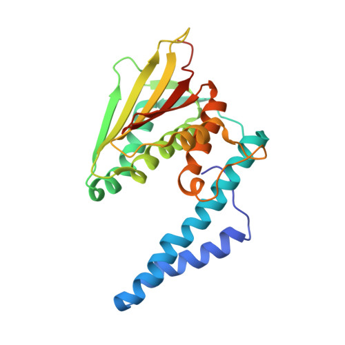
    Recent paradigm shifting discoveries have demonstrated that bacterial signaling kinases engage in unexpected regulatory crosstalk, yet the underlying molecular mechanisms remain largely uncharacterized. The Pseudomonas aeruginosa RetS/GacS system constitutes an ideal model for studying these mechanisms. The in-depth analysis of the kinase region of RetS and RetS/GacS interactions presented here refutes a longstanding model, which posited the formation of a catalytically inactive RetS/GacS heterodimer. Crystallographic studies uncovered structurally dynamic features within the RetS kinase region, suggesting that RetS uses the reversible unfolding of a helix, or helix cracking, to control interactions with GacS. The pivotal importance of this helical region for regulating GacS and, by extension, Pseudomonas aeruginosa virulence, was corroborated via in vivo assays. The implications of this work extend beyond the RetS/GacS system because the helix cracking occurs right next to a highly conserved catalytic residue histidine-424, suggesting this model could represent an emergent archetype for histidine kinase regulation.


  • Organizational Affiliation

    Department of Biological Sciences, Virginia Polytechnic Institute and State University, Blacksburg, VA 24061, USA.


Macromolecules
Find similar proteins by:  (by identity cutoff)  |  3D Structure
Entity ID: 1
MoleculeChains Sequence LengthOrganismDetailsImage
RetS (Regulator of Exopolysaccharide and Type III Secretion)
A, B, C, D, E
A, B, C, D, E, F, G, H
234Pseudomonas aeruginosaMutation(s): 0 
Gene Names: retSPA4856
UniProt
Find proteins for Q9HUV7 (Pseudomonas aeruginosa (strain ATCC 15692 / DSM 22644 / CIP 104116 / JCM 14847 / LMG 12228 / 1C / PRS 101 / PAO1))
Explore Q9HUV7 
Go to UniProtKB:  Q9HUV7
Entity Groups  
Sequence Clusters30% Identity50% Identity70% Identity90% Identity95% Identity100% Identity
UniProt GroupQ9HUV7
Sequence Annotations
Expand
  • Reference Sequence
Small Molecules
Ligands 1 Unique
IDChains Name / Formula / InChI Key2D Diagram3D Interactions
NI
Query on NI

Download Ideal Coordinates CCD File 
AA [auth H]
BA [auth H]
I [auth A]
J [auth A]
K [auth A]
AA [auth H],
BA [auth H],
I [auth A],
J [auth A],
K [auth A],
L [auth B],
M [auth B],
N [auth B],
O [auth C],
P [auth C],
Q [auth C],
R [auth D],
S [auth E],
T [auth E],
U [auth E],
V [auth F],
W [auth F],
X [auth G],
Y [auth G],
Z [auth G]
NICKEL (II) ION
Ni
VEQPNABPJHWNSG-UHFFFAOYSA-N
Experimental Data & Validation

Experimental Data

  • Method: X-RAY DIFFRACTION
  • Resolution: 3.80 Å
  • R-Value Free: 0.249 
  • R-Value Work: 0.215 
  • R-Value Observed: 0.215 
  • Space Group: P 41 21 2
Unit Cell:
Length ( Å )Angle ( ˚ )
a = 158.708α = 90
b = 158.708β = 90
c = 243.826γ = 90
Software Package:
Software NamePurpose
PHENIXrefinement
PDB_EXTRACTdata extraction
MOSFLMdata reduction
SCALAdata scaling
PHENIXphasing

Structure Validation

View Full Validation Report


Entry History 

Deposition Data

Revision History  (Full details and data files)

  • Version 1.0: 2019-03-20
    Type: Initial release
  • Version 1.1: 2019-04-03
    Changes: Data collection, Database references
  • Version 1.2: 2019-04-17
    Changes: Author supporting evidence, Data collection
  • Version 1.3: 2019-05-22
    Changes: Data collection, Database references
  • Version 1.4: 2023-10-11
    Changes: Data collection, Database references, Derived calculations, Refinement description