6DJX

Crystal Structure of pParkin-pUb-UbcH7 complex


Experimental Data Snapshot

  • Method: X-RAY DIFFRACTION
  • Resolution: 4.80 Å
  • R-Value Free: 0.290 
  • R-Value Work: 0.257 
  • R-Value Observed: 0.259 

wwPDB Validation   3D Report Full Report


This is version 1.4 of the entry. See complete history


Literature

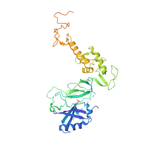
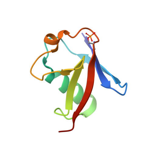
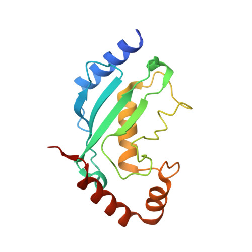
Mechanism of parkin activation by phosphorylation.

Sauve, V.Sung, G.Soya, N.Kozlov, G.Blaimschein, N.Miotto, L.S.Trempe, J.F.Lukacs, G.L.Gehring, K.

(2018) Nat Struct Mol Biol 25: 623-630

  • DOI: https://doi.org/10.1038/s41594-018-0088-7
  • Primary Citation of Related Structures:  
    6DJW, 6DJX

  • PubMed Abstract: 

    Mutations in the ubiquitin ligase parkin are responsible for a familial form of Parkinson's disease. Parkin and the PINK1 kinase regulate a quality-control system for mitochondria. PINK1 phosphorylates ubiquitin on the outer membrane of damaged mitochondria, thus leading to recruitment and activation of parkin via phosphorylation of its ubiquitin-like (Ubl) domain. Here, we describe the mechanism of parkin activation by phosphorylation. The crystal structure of phosphorylated Bactrocera dorsalis (oriental fruit fly) parkin in complex with phosphorylated ubiquitin and an E2 ubiquitin-conjugating enzyme reveals that the key activating step is movement of the Ubl domain and release of the catalytic RING2 domain. Hydrogen/deuterium exchange and NMR experiments with the various intermediates in the activation pathway confirm and extend the interpretation of the crystal structure to mammalian parkin. Our results rationalize previously unexplained Parkinson's disease mutations and the presence of internal linkers that allow large domain movements in parkin.


  • Organizational Affiliation

    Department of Biochemistry, McGill University, Montreal, Quebec, Canada.


Macromolecules
Find similar proteins by:  (by identity cutoff)  |  3D Structure
Entity ID: 1
MoleculeChains Sequence LengthOrganismDetailsImage
RBR-type E3 ubiquitin transferase,RBR-type E3 ubiquitin transferase432Bactrocera dorsalisMutation(s): 1 
Gene Names: PRKN2
EC: 2.3.2.31
UniProt
Find proteins for A0A034W4L8 (Bactrocera dorsalis)
Explore A0A034W4L8 
Go to UniProtKB:  A0A034W4L8
Entity Groups  
Sequence Clusters30% Identity50% Identity70% Identity90% Identity95% Identity100% Identity
UniProt GroupA0A034W4L8
Sequence Annotations
Expand
  • Reference Sequence
Find similar proteins by:  (by identity cutoff)  |  3D Structure
Entity ID: 2
MoleculeChains Sequence LengthOrganismDetailsImage
Ubiquitin76Bos taurusMutation(s): 0 
UniProt
Find proteins for P62992 (Bos taurus)
Explore P62992 
Go to UniProtKB:  P62992
Entity Groups  
Sequence Clusters30% Identity50% Identity70% Identity90% Identity95% Identity100% Identity
UniProt GroupP62992
Sequence Annotations
Expand
  • Reference Sequence
Find similar proteins by:  (by identity cutoff)  |  3D Structure
Entity ID: 3
MoleculeChains Sequence LengthOrganismDetailsImage
Ubiquitin-conjugating enzyme E2 L3159Homo sapiensMutation(s): 1 
Gene Names: UBE2L3UBCE7UBCH7
EC: 2.3.2.23
UniProt & NIH Common Fund Data Resources
Find proteins for P68036 (Homo sapiens)
Explore P68036 
Go to UniProtKB:  P68036
PHAROS:  P68036
GTEx:  ENSG00000185651 
Entity Groups  
Sequence Clusters30% Identity50% Identity70% Identity90% Identity95% Identity100% Identity
UniProt GroupP68036
Sequence Annotations
Expand
  • Reference Sequence
Experimental Data & Validation

Experimental Data

  • Method: X-RAY DIFFRACTION
  • Resolution: 4.80 Å
  • R-Value Free: 0.290 
  • R-Value Work: 0.257 
  • R-Value Observed: 0.259 
  • Space Group: P 31 2 1
Unit Cell:
Length ( Å )Angle ( ˚ )
a = 135.669α = 90
b = 135.669β = 90
c = 87.993γ = 120
Software Package:
Software NamePurpose
XDSdata reduction
Aimlessdata scaling
PHENIXrefinement
PDB_EXTRACTdata extraction
PHASERphasing

Structure Validation

View Full Validation Report


Entry History & Funding Information

Deposition Data


Funding OrganizationLocationGrant Number
Canadian Institutes of Health Research (CIHR)CanadaMOP-125924
Michael J. Fox FoundationUnited States12144

Revision History  (Full details and data files)

  • Version 1.0: 2018-07-04
    Type: Initial release
  • Version 1.1: 2018-07-18
    Changes: Data collection, Database references
  • Version 1.2: 2018-07-25
    Changes: Data collection, Database references
  • Version 1.3: 2020-01-08
    Changes: Author supporting evidence
  • Version 1.4: 2023-10-11
    Changes: Data collection, Database references, Derived calculations, Refinement description