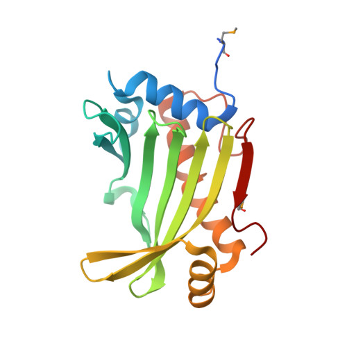
6DHX | pdb_00006dhx

Structure of TipC2 from Streptococcus intermedius B196


Experimental Data Snapshot

  • Method: X-RAY DIFFRACTION
  • Resolution: 1.75 Å
  • R-Value Free: 
    0.194 (Depositor), 0.195 (DCC) 
  • R-Value Work: 
    0.170 (Depositor), 0.171 (DCC) 
  • R-Value Observed: 
    0.171 (Depositor) 

wwPDB Validation 3D Report Full Report

Validation slider image for 6DHX

This is version 1.3 of the entry. See complete history

Literature

Molecular Basis for Immunity Protein Recognition of a Type VII Secretion System Exported Antibacterial Toxin.

Klein, T.A.Pazos, M.Surette, M.G.Vollmer, W.Whitney, J.C.

(2018) J Mol Biology 430: 4344-4358

  • DOI: https://doi.org/10.1016/j.jmb.2018.08.027
  • Primary Citation Related Structures: 
    6DHX

  • PubMed Abstract: 

    Gram-positive bacteria deploy the type VII secretion system (T7SS) to facilitate interactions between eukaryotic and prokaryotic cells. In recent work, we identified the TelC protein from Streptococcus intermedius as a T7SS-exported lipid II phosphatase that mediates interbacterial competition. TelC exerts toxicity in the inner wall zone of Gram-positive bacteria; however, intercellular intoxication of sister cells does not occur because they express the TipC immunity protein. In the present study, we sought to characterize the molecular basis of self-protection by TipC. Using sub-cellular localization and protease protection assays, we show that TipC is a membrane protein with an N-terminal transmembrane segment and a C-terminal TelC-inhibitory domain that protrudes into the inner wall zone. The 1.9-Å X-ray crystal structure of a non-protective TipC paralogue reveals that the soluble domain of TipC proteins adopts a crescent-shaped fold that is composed of three α-helices and a seven-stranded β-sheet. Subsequent homology-guided mutagenesis demonstrates that a concave surface formed by the predicted β-sheet of TipC is required for both its interaction with TelC and its TelC-inhibitory activity. S. intermedius cells lacking the tipC gene are susceptible to growth inhibition by TelC delivered between cells; however, we find that the growth of this strain is unaffected by endogenous or overexpressed TelC, although the toxin accumulates in culture supernatants. Together, these data indicate that the TelC-inhibitory activity of TipC is only required for intercellularly transferred TelC and that the T7SS apparatus transports TelC across the cell envelope in a single step, bypassing the cellular compartment in which it exerts toxicity en route.


  • Organizational Affiliation
    • Michael DeGroote Institute for Infectious Disease Research, McMaster University, Hamilton, Canada L8S 4K1; Department of Biochemistry and Biomedical Sciences, McMaster University, Hamilton, Canada L8S 4L8.

Macromolecule Content 

  • Total Structure Weight: 70.64 kDa 
  • Atom Count: 4,978 
  • Modeled Residue Count: 539 
  • Deposited Residue Count: 585 
  • Unique protein chains: 1

Macromolecules

Find similar proteins by:|  3D Structure
Entity ID: 1
MoleculeChains  Sequence LengthOrganismDetailsImage
TipC2
A, B, C
195Streptococcus intermedius B196Mutation(s): 0 
Gene Names: SIR_1486
UniProt
Find proteins for T1ZH71 (Streptococcus intermedius B196)
Explore T1ZH71 
Go to UniProtKB:  T1ZH71
Entity Groups
Sequence Clusters30% Identity50% Identity70% Identity90% Identity95% Identity100% Identity
UniProt GroupT1ZH71
Sequence Annotations
Expand
Reference Sequence

Small Molecules

Ligands 1 Unique
IDChains Name / Formula / InChI Key2D Diagram3D Interactions
SO4

Query on SO4


Download:Ideal Coordinates CCD File
D [auth A]SULFATE ION
O4 S
QAOWNCQODCNURD-UHFFFAOYSA-L
Modified Residues  1 Unique
IDChains TypeFormula2D DiagramParent
MSE
Query on MSE
A, B, C
L-PEPTIDE LINKINGC5 H11 N O2 SeMET

Experimental Data & Validation

Experimental Data

  • Method: X-RAY DIFFRACTION
  • Resolution: 1.75 Å
  • R-Value Free:  0.194 (Depositor), 0.195 (DCC) 
  • R-Value Work:  0.170 (Depositor), 0.171 (DCC) 
  • R-Value Observed: 0.171 (Depositor) 
Space Group: C 1 2 1
Unit Cell:
Length ( Å )Angle ( ˚ )
a = 159.74α = 90
b = 54.52β = 108.05
c = 104.43γ = 90
Software Package:
Software NamePurpose
PHENIXrefinement
xia2data reduction
Aimlessdata scaling
PHENIXphasing

Structure Validation

View Full Validation Report


Entry History 

& Funding Information

Deposition Data


Funding OrganizationLocationGrant Number
National Institutes of Health/National Institute Of Allergy and Infectious Diseases (NIH/NIAID)United StatesAI080609

Revision History  (Full details and data files)

  • Version 1.0: 2018-09-19
    Type: Initial release
  • Version 1.1: 2018-10-24
    Changes: Data collection, Database references
  • Version 1.2: 2019-12-18
    Changes: Author supporting evidence
  • Version 1.3: 2024-10-30
    Changes: Data collection, Database references, Structure summary