A Cell-Surface GH9 Endo-Glucanase Coordinates with Surface Glycan-Binding Proteins to Mediate Xyloglucan Uptake in the Gut Symbiont Bacteroides ovatus.
Foley, M.H., Dejean, G., Hemsworth, G.R., Davies, G.J., Brumer, H., Koropatkin, N.M.(2019) J Mol Biology 431: 981-995
- PubMed: 30668971 Search on PubMedSearch on PubMed Central
- DOI: https://doi.org/10.1016/j.jmb.2019.01.008
- Primary Citation Related Structures:
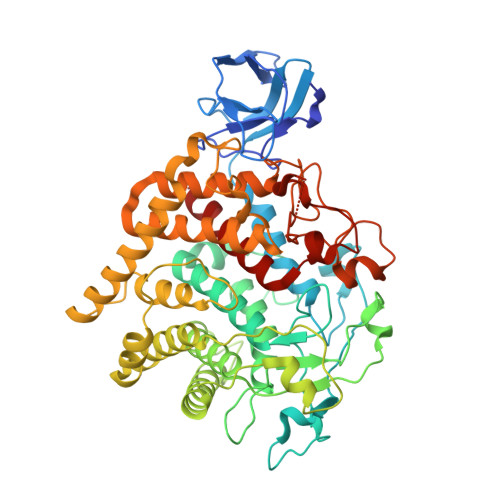
6DHT - PubMed Abstract:
Dietary fiber is an important food source for members of the human gut microbiome. Members of the dominant Bacteroidetes phylum capture diverse polysaccharides via the action of multiple cell surface proteins encoded within polysaccharide utilization loci (PUL). The independent activities of PUL-encoded glycoside hydrolases (GHs) and surface glycan-binding proteins (SGBPs) for the harvest of various glycans have been studied in detail, but how these proteins work together to coordinate uptake is poorly understood. Here, we combine genetic and biochemical approaches to discern the interplay between the BoGH9 endoglucanase and the xyloglucan-binding proteins SGBP-A and SGBP-B from the Bacteroides ovatus xyloglucan utilization locus (XyGUL). The expression of BoGH9, a weakly active xyloglucanase in isolation, is required in a strain that expresses a non-binding version of SGBP-A (SGBP-A*). The crystal structure of the BoGH9 enzyme suggests the molecular basis for its robust activity on mixed-linkage β-glucan compared to xyloglucan. However, catalytically inactive site-directed mutants of BoGH9 fail to complement the deletion of the active BoGH9 in a SGBP-A* strain. We also find that SGBP-B is needed in an SGBP-A* background to support growth on xyloglucan, but that the non-binding SGBP-B* protein acts in a dominant negative manner to inhibit growth on xyloglucan. We postulate a model whereby the SGBP-A, SGBP-B, and BoGH9 work together at the cell surface, likely within a discrete complex, and that xyloglucan binding by SGBP-B and BoGH9 may facilitate the orientation of the xyloglucan for transfer across the outer membrane.
- Department of Microbiology and Immunology, University of Michigan Medical School, Ann Arbor, MI 48109, USA.
Organizational Affiliation: