6DFY | pdb_00006dfy

Remodeled crystal structure of DNA-bound DUX4-HD2


Experimental Data Snapshot

  • Method: X-RAY DIFFRACTION
  • Resolution: 2.62 Å
  • R-Value Free: 
    0.279 (Depositor), 0.283 (DCC) 
  • R-Value Work: 
    0.246 (Depositor), 0.253 (DCC) 
  • R-Value Observed: 
    0.247 (Depositor) 

Starting Model: experimental
View more details

wwPDB Validation 3D Report Full Report

Validation slider image for 6DFY

This is version 1.3 of the entry. See complete history

Literature

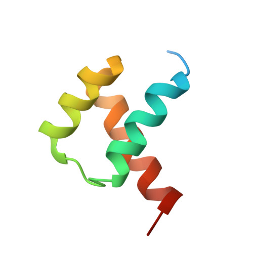
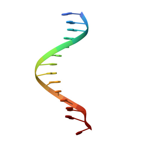
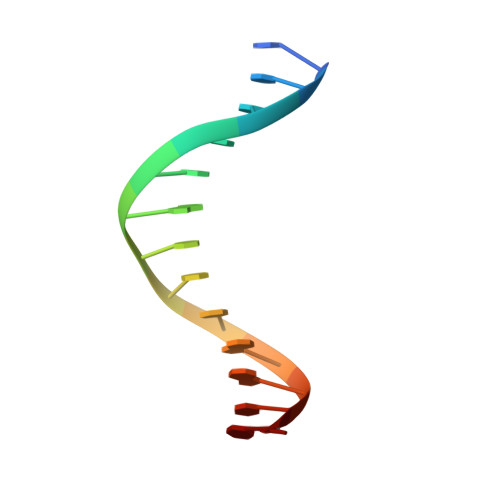
Comment on structural basis of DUX4/IGH-driven transactivation.

Aihara, H.Shi, K.Lee, J.K.Bosnakovski, D.Kyba, M.

(2018) Leukemia 32: 2090-2092

  • DOI: https://doi.org/10.1038/s41375-018-0217-7
  • Primary Citation Related Structures: 
    6DFY

  • Organizational Affiliation
    • Department of Biochemistry, Molecular Biology and Biophysics, University of Minnesota, Minneapolis, MN, 55455, USA. aihar001@umn.edu.

Macromolecule Content 

  • Total Structure Weight: 23.13 kDa 
  • Atom Count: 1,420 
  • Modeled Residue Count: 134 
  • Deposited Residue Count: 156 
  • Unique protein chains: 1
  • Unique nucleic acid chains: 2

Macromolecules


Find similar proteins by:|  3D Structure
Entity ID: 1
MoleculeChains  Sequence LengthOrganismDetailsImage
Double homeobox protein 4A [auth C],
D
64Homo sapiensMutation(s): 0 
Gene Names: DUX4DUX10
UniProt & NIH Common Fund Data Resources
Find proteins for Q9UBX2 (Homo sapiens)
Explore Q9UBX2 
Go to UniProtKB:  Q9UBX2
PHAROS:  Q9UBX2
GTEx:  ENSG00000260596 
Entity Groups
Sequence Clusters30% Identity50% Identity70% Identity90% Identity95% Identity100% Identity
UniProt GroupQ9UBX2
Sequence Annotations
Expand
Reference Sequence
Find similar nucleic acids by:  Sequence
Entity ID: 2
MoleculeChains LengthOrganismImage
DNA (5'-D(*AP*AP*GP*AP*TP*TP*AP*GP*AP*TP*TP*AP*GP*T)-3')B [auth E]14synthetic construct
Sequence Annotations
Expand
Reference Sequence
Find similar nucleic acids by:  Sequence
Entity ID: 3
MoleculeChains LengthOrganismImage
DNA (5'-D(*TP*TP*CP*TP*AP*AP*TP*CP*TP*AP*AP*TP*CP*A)-3')C [auth F]14synthetic construct
Sequence Annotations
Expand
Reference Sequence

Experimental Data & Validation

Experimental Data

  • Method: X-RAY DIFFRACTION
  • Resolution: 2.62 Å
  • R-Value Free:  0.279 (Depositor), 0.283 (DCC) 
  • R-Value Work:  0.246 (Depositor), 0.253 (DCC) 
  • R-Value Observed: 0.247 (Depositor) 
Space Group: P 41 21 2
Unit Cell:
Length ( Å )Angle ( ˚ )
a = 51.61α = 90
b = 51.61β = 90
c = 166.55γ = 90
Software Package:
Software NamePurpose
PHENIXrefinement
MOSFLMdata reduction
SCALAdata scaling
PHENIXphasing

Structure Validation

View Full Validation Report


Entry History 

& Funding Information

Deposition Data


Funding OrganizationLocationGrant Number
National Institutes of Health/National Institute of General Medical Sciences (NIH/NIGMS)United StatesGM103403

Revision History  (Full details and data files)

  • Version 1.0: 2018-09-05
    Type: Initial release
  • Version 1.1: 2018-09-19
    Changes: Data collection, Database references
  • Version 1.2: 2020-01-01
    Changes: Author supporting evidence
  • Version 1.3: 2023-10-11
    Changes: Data collection, Database references, Refinement description