6D8K | pdb_00006d8k

Bacteroides multiple species beta-glucuronidase


Experimental Data Snapshot

  • Method: X-RAY DIFFRACTION
  • Resolution: 2.65 Å
  • R-Value Free: 
    0.283 (Depositor), 0.280 (DCC) 
  • R-Value Work: 
    0.207 (Depositor) 
  • R-Value Observed: 
    0.209 (Depositor) 

wwPDB Validation 3D Report Full Report

Validation slider image for 6D8K

This is version 1.4 of the entry. See complete history

Literature

Three structurally and functionally distinct beta-glucuronidases from the human gut microbeBacteroides uniformis.

Pellock, S.J.Walton, W.G.Biernat, K.A.Torres-Rivera, D.Creekmore, B.C.Xu, Y.Liu, J.Tripathy, A.Stewart, L.J.Redinbo, M.R.

(2018) J Biological Chem 293: 18559-18573

  • DOI: https://doi.org/10.1074/jbc.RA118.005414
  • Primary Citation Related Structures: 
    6D1N, 6D1P, 6D41, 6D50, 6D6W, 6D7F, 6D89, 6D8G, 6D8K

  • PubMed Abstract: 

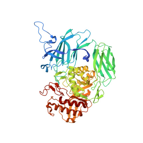
    The glycoside hydrolases encoded by the human gut microbiome play an integral role in processing a variety of exogenous and endogenous glycoconjugates. Here we present three structurally and functionally distinct β-glucuronidase (GUS) glycoside hydrolases from a single human gut commensal microbe, Bacteroides uniformis We show using nine crystal structures, biochemical, and biophysical data that whereas these three proteins share similar overall folds, they exhibit different structural features that create three structurally and functionally unique enzyme active sites. Notably, quaternary structure plays an important role in creating distinct active site features that are hard to predict via structural modeling methods. The enzymes display differential processing capabilities toward glucuronic acid-containing polysaccharides and SN-38-glucuronide, a metabolite of the cancer drug irinotecan. We also demonstrate that GUS-specific and nonselective inhibitors exhibit varying potencies toward each enzyme. Together, these data highlight the diversity of GUS enzymes within a single Bacteroides gut commensal and advance our understanding of how structural details impact the specific roles microbial enzymes play in processing drug-glucuronide and glycan substrates.


  • Organizational Affiliation
    • From the Departments of Chemistry.

Macromolecule Content 

  • Total Structure Weight: 277.39 kDa 
  • Atom Count: 17,233 
  • Modeled Residue Count: 2,085 
  • Deposited Residue Count: 2,388 
  • Unique protein chains: 1

Macromolecules

Find similar proteins by:|  3D Structure
Entity ID: 1
MoleculeChains  Sequence LengthOrganismDetailsImage
Glycosyl hydrolase family 2, sugar binding domain protein
A, B, C, D
597Bacteroides ovatusMutation(s): 0 
Gene Names: HMPREF2532_04191

Experimental Data & Validation

Experimental Data

  • Method: X-RAY DIFFRACTION
  • Resolution: 2.65 Å
  • R-Value Free:  0.283 (Depositor), 0.280 (DCC) 
  • R-Value Work:  0.207 (Depositor) 
  • R-Value Observed: 0.209 (Depositor) 
Space Group: I 41
Unit Cell:
Length ( Å )Angle ( ˚ )
a = 213.464α = 90
b = 213.464β = 90
c = 112.607γ = 90
Software Package:
Software NamePurpose
PHENIXrefinement
XDSdata reduction
Aimlessdata scaling
PHENIXphasing

Structure Validation

View Full Validation Report


Entry History 

& Funding Information

Deposition Data


Funding OrganizationLocationGrant Number
National Institutes of Health/National Cancer Institute (NIH/NCI)United StatesCA098468
National Institutes of Health/National Cancer Institute (NIH/NCI)United StatesCA207416

Revision History  (Full details and data files)

  • Version 1.0: 2018-10-17
    Type: Initial release
  • Version 1.1: 2018-10-24
    Changes: Data collection, Database references
  • Version 1.2: 2018-12-19
    Changes: Data collection, Database references
  • Version 1.3: 2019-12-04
    Changes: Author supporting evidence
  • Version 1.4: 2024-03-13
    Changes: Data collection, Database references