6D6T

Human GABA-A receptor alpha1-beta2-gamma2 subtype in complex with GABA and flumazenil, conformation B


Experimental Data Snapshot

  • Method: ELECTRON MICROSCOPY
  • Resolution: 3.86 Å
  • Aggregation State: PARTICLE 
  • Reconstruction Method: SINGLE PARTICLE 

wwPDB Validation   3D Report Full Report


This is version 3.0 of the entry. See complete history


Literature

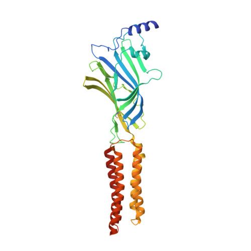
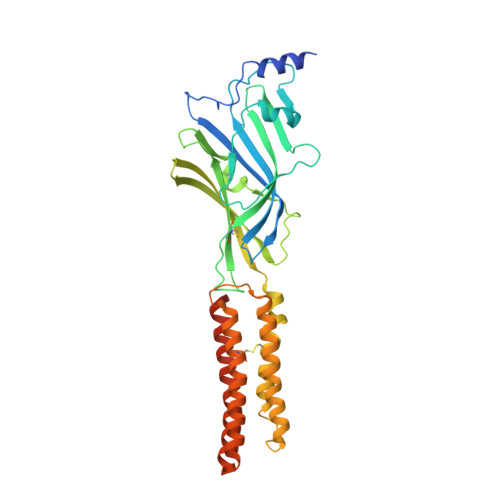
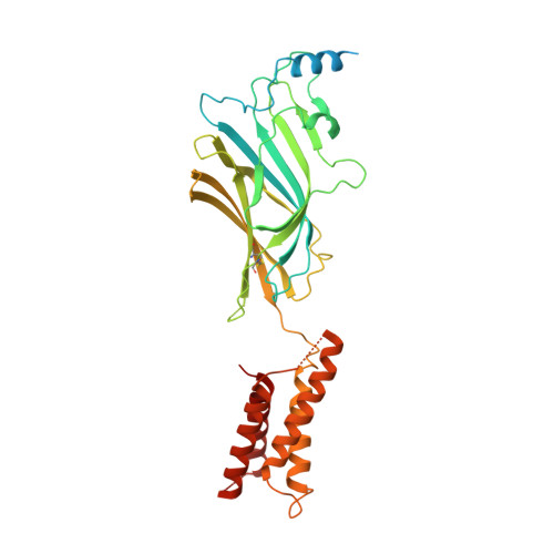
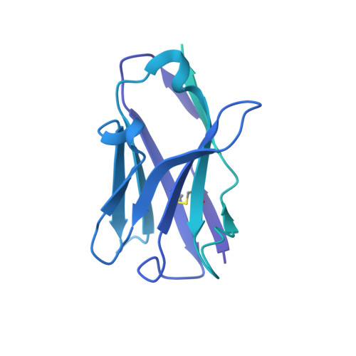
Structure of a human synaptic GABAAreceptor.

Zhu, S.Noviello, C.M.Teng, J.Walsh, R.M.Kim, J.J.Hibbs, R.E.

(2018) Nature 559: 67-72

  • DOI: https://doi.org/10.1038/s41586-018-0255-3
  • Primary Citation of Related Structures:  
    6D6T, 6D6U

  • PubMed Abstract: 

    Fast inhibitory neurotransmission in the brain is principally mediated by the neurotransmitter GABA (γ-aminobutyric acid) and its synaptic target, the type A GABA receptor (GABA A receptor). Dysfunction of this receptor results in neurological disorders and mental illnesses including epilepsy, anxiety and insomnia. The GABA A receptor is also a prolific target for therapeutic, illicit and recreational drugs, including benzodiazepines, barbiturates, anaesthetics and ethanol. Here we present high-resolution cryo-electron microscopy structures of the human α1β2γ2 GABA A receptor, the predominant isoform in the adult brain, in complex with GABA and the benzodiazepine site antagonist flumazenil, the first-line clinical treatment for benzodiazepine overdose. The receptor architecture reveals unique heteromeric interactions for this important class of inhibitory neurotransmitter receptor. This work provides a template for understanding receptor modulation by GABA and benzodiazepines, and will assist rational approaches to therapeutic targeting of this receptor for neurological disorders and mental illness.


  • Organizational Affiliation

    Departments of Neuroscience and Biophysics, University of Texas Southwestern Medical Center, Dallas, TX, USA.


Macromolecules
Find similar proteins by:  (by identity cutoff)  |  3D Structure
Entity ID: 1
MoleculeChains Sequence LengthOrganismDetailsImage
Gamma-aminobutyric acid receptor subunit beta-2,Gamma-aminobutyric acid receptor subunit beta-2
A, C
341Homo sapiensMutation(s): 0 
Gene Names: GABRB2
Membrane Entity: Yes 
UniProt & NIH Common Fund Data Resources
Find proteins for P47870 (Homo sapiens)
Explore P47870 
Go to UniProtKB:  P47870
PHAROS:  P47870
GTEx:  ENSG00000145864 
Entity Groups  
Sequence Clusters30% Identity50% Identity70% Identity90% Identity95% Identity100% Identity
UniProt GroupP47870
Sequence Annotations
Expand
  • Reference Sequence
Find similar proteins by:  (by identity cutoff)  |  3D Structure
Entity ID: 2
MoleculeChains Sequence LengthOrganismDetailsImage
Gamma-aminobutyric acid receptor subunit alpha-1,Gamma-aminobutyric acid receptor subunit alpha-1
B, D
358Homo sapiensMutation(s): 0 
Gene Names: GABRA1
Membrane Entity: Yes 
UniProt & NIH Common Fund Data Resources
Find proteins for P14867 (Homo sapiens)
Explore P14867 
Go to UniProtKB:  P14867
PHAROS:  P14867
GTEx:  ENSG00000022355 
Entity Groups  
Sequence Clusters30% Identity50% Identity70% Identity90% Identity95% Identity100% Identity
UniProt GroupP14867
Sequence Annotations
Expand
  • Reference Sequence
Find similar proteins by:  (by identity cutoff)  |  3D Structure
Entity ID: 3
MoleculeChains Sequence LengthOrganismDetailsImage
Human GABA-A receptor subunit gamma-2366Homo sapiensMutation(s): 0 
Gene Names: GABRG2
Membrane Entity: Yes 
UniProt & NIH Common Fund Data Resources
Find proteins for P18507 (Homo sapiens)
Explore P18507 
Go to UniProtKB:  P18507
PHAROS:  P18507
GTEx:  ENSG00000113327 
Entity Groups  
Sequence Clusters30% Identity50% Identity70% Identity90% Identity95% Identity100% Identity
UniProt GroupP18507
Sequence Annotations
Expand
  • Reference Sequence
Find similar proteins by:  (by identity cutoff)  |  3D Structure
Entity ID: 4
MoleculeChains Sequence LengthOrganismDetailsImage
Kappa Fab Light ChainF [auth I],
H [auth L]
213Mus musculusMutation(s): 0 
Entity Groups  
Sequence Clusters30% Identity50% Identity70% Identity90% Identity95% Identity100% Identity
Sequence Annotations
Expand
  • Reference Sequence
Find similar proteins by:  (by identity cutoff)  |  3D Structure
Entity ID: 5
MoleculeChains Sequence LengthOrganismDetailsImage
IgG2b Fab Heavy ChainG [auth J],
I [auth K]
454Mus musculusMutation(s): 0 
Entity Groups  
Sequence Clusters30% Identity50% Identity70% Identity90% Identity95% Identity100% Identity
Sequence Annotations
Expand
  • Reference Sequence
Oligosaccharides

Help

Entity ID: 6
MoleculeChains Length2D Diagram Glycosylation3D Interactions
beta-D-mannopyranose-(1-4)-2-acetamido-2-deoxy-beta-D-glucopyranose-(1-4)-2-acetamido-2-deoxy-beta-D-glucopyranoseJ [auth F],
L [auth H]
3N-Glycosylation
Glycosylation Resources
GlyTouCan:  G15407YE
GlyCosmos:  G15407YE
GlyGen:  G15407YE
Entity ID: 7
MoleculeChains Length2D Diagram Glycosylation3D Interactions
alpha-D-mannopyranose-(1-2)-alpha-D-mannopyranose-(1-3)-[alpha-D-mannopyranose-(1-3)-[alpha-D-mannopyranose-(1-6)]alpha-D-mannopyranose-(1-6)]beta-D-mannopyranose-(1-4)-2-acetamido-2-deoxy-beta-D-glucopyranose-(1-4)-2-acetamido-2-deoxy-beta-D-glucopyranoseK [auth G]8N-Glycosylation
Glycosylation Resources
GlyTouCan:  G80966KZ
GlyCosmos:  G80966KZ
GlyGen:  G80966KZ
Entity ID: 8
MoleculeChains Length2D Diagram Glycosylation3D Interactions
alpha-D-mannopyranose-(1-3)-[alpha-D-mannopyranose-(1-6)]beta-D-mannopyranose-(1-4)-2-acetamido-2-deoxy-beta-D-glucopyranose-(1-4)-2-acetamido-2-deoxy-beta-D-glucopyranose
M
5N-Glycosylation
Glycosylation Resources
GlyTouCan:  G22768VO
GlyCosmos:  G22768VO
GlyGen:  G22768VO
Small Molecules
Ligands 4 Unique
IDChains Name / Formula / InChI Key2D Diagram3D Interactions
Y01
Query on Y01

Download Ideal Coordinates CCD File 
BA [auth D]
CA [auth D]
DA [auth D]
P [auth A]
Q [auth A]
BA [auth D],
CA [auth D],
DA [auth D],
P [auth A],
Q [auth A],
R [auth A],
S [auth A],
T [auth A],
U [auth B],
V [auth B],
W [auth B],
X [auth B]
CHOLESTEROL HEMISUCCINATE
C31 H50 O4
WLNARFZDISHUGS-MIXBDBMTSA-N
FYP
Query on FYP

Download Ideal Coordinates CCD File 
AA [auth D]ethyl 8-fluoro-5-methyl-6-oxo-5,6-dihydro-4H-imidazo[1,5-a][1,4]benzodiazepine-3-carboxylate
C15 H14 F N3 O3
OFBIFZUFASYYRE-UHFFFAOYSA-N
NAG
Query on NAG

Download Ideal Coordinates CCD File 
EA [auth E],
N [auth A],
Y [auth C]
2-acetamido-2-deoxy-beta-D-glucopyranose
C8 H15 N O6
OVRNDRQMDRJTHS-FMDGEEDCSA-N
ABU
Query on ABU

Download Ideal Coordinates CCD File 
O [auth A],
Z [auth C]
GAMMA-AMINO-BUTANOIC ACID
C4 H9 N O2
BTCSSZJGUNDROE-UHFFFAOYSA-N
Binding Affinity Annotations 
IDSourceBinding Affinity
FYP BindingDB:  6D6T Ki: min: 0.19, max: 148 (nM) from 45 assay(s)
Kd: 0.45 (nM) from 1 assay(s)
ABU BindingDB:  6D6T Ki: min: 21.37, max: 77.62 (nM) from 4 assay(s)
IC50: 22 (nM) from 1 assay(s)
EC50: min: 650, max: 8.00e+4 (nM) from 11 assay(s)
Experimental Data & Validation

Experimental Data

  • Method: ELECTRON MICROSCOPY
  • Resolution: 3.86 Å
  • Aggregation State: PARTICLE 
  • Reconstruction Method: SINGLE PARTICLE 
EM Software:
TaskSoftware PackageVersion
RECONSTRUCTIONRELION2.1
MODEL REFINEMENTPHENIX1.13-2988

Structure Validation

View Full Validation Report


Entry History & Funding Information

Deposition Data


Funding OrganizationLocationGrant Number
Welch FoundationUnited StatesI-1812

Revision History  (Full details and data files)

  • Version 1.0: 2018-06-27
    Type: Initial release
  • Version 1.1: 2018-07-11
    Changes: Data collection, Database references
  • Version 1.2: 2018-07-18
    Changes: Data collection, Database references
  • Version 1.3: 2019-12-18
    Changes: Other
  • Version 1.4: 2020-07-01
    Changes: Derived calculations, Structure summary
  • Version 2.0: 2020-07-29
    Type: Remediation
    Reason: Carbohydrate remediation
    Changes: Advisory, Atomic model, Data collection, Derived calculations, Structure summary
  • Version 3.0: 2023-11-15
    Changes: Atomic model, Data collection, Database references, Refinement description, Structure summary