6D3B

INFLUENZA VIRUS NEURAMINIDASE SUBTYPE N9 (TERN) APO FORM


Experimental Data Snapshot

  • Method: X-RAY DIFFRACTION
  • Resolution: 1.40 Å
  • R-Value Free: 0.170 
  • R-Value Work: 0.152 
  • R-Value Observed: 0.153 

wwPDB Validation   3D Report Full Report


Ligand Structure Quality Assessment 


This is version 2.1 of the entry. See complete history


Literature

Structure of an Influenza A virus N9 neuraminidase with a tetrabrachion-domain stalk.

Streltsov, V.A.Schmidt, P.M.McKimm-Breschkin, J.L.

(2019) Acta Crystallogr F Struct Biol Commun 75: 89-97

  • DOI: https://doi.org/10.1107/S2053230X18017892
  • Primary Citation of Related Structures:  
    6CRD, 6D3B, 6MCX

  • PubMed Abstract: 

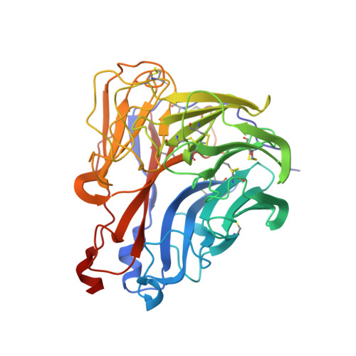
    The influenza neuraminidase (NA) is a homotetramer with head, stalk, transmembrane and cytoplasmic regions. The structure of the NA head with a stalk has never been determined. The NA head from an N9 subtype influenza A virus, A/tern/Australia/G70C/1975 (H1N9), was expressed with an artificial stalk derived from the tetrabrachion (TB) tetramerization domain from Staphylothermus marinus. The NA was successfully crystallized both with and without the TB stalk, and the structures were determined to 2.6 and 2.3 Å resolution, respectively. Comparisons of the two NAs with the native N9 NA structure from egg-grown virus showed that the artificial TB stalk maintained the native NA head structure, supporting previous biological observations.


  • Organizational Affiliation

    CSIRO Manufacturing, 343 Royal Parade, Parkville, Victoria 3052, Australia.


Macromolecules
Find similar proteins by:  (by identity cutoff)  |  3D Structure
Entity ID: 1
MoleculeChains Sequence LengthOrganismDetailsImage
Neuraminidase389Influenza A virus (A/tern/Australia/G70C/1975(H11N9))Mutation(s): 0 
EC: 3.2.1.18
UniProt
Find proteins for P03472 (Influenza A virus (strain A/Tern/Australia/G70C/1975 H11N9))
Explore P03472 
Go to UniProtKB:  P03472
Entity Groups  
Sequence Clusters30% Identity50% Identity70% Identity90% Identity95% Identity100% Identity
UniProt GroupP03472
Sequence Annotations
Expand
  • Reference Sequence
Oligosaccharides

Help

Entity ID: 2
MoleculeChains Length2D Diagram Glycosylation3D Interactions
alpha-D-mannopyranose-(1-2)-alpha-D-mannopyranose-(1-2)-alpha-D-mannopyranose-(1-3)-[alpha-D-mannopyranose-(1-3)-[alpha-D-mannopyranose-(1-6)]alpha-D-mannopyranose-(1-6)]beta-D-mannopyranose-(1-4)-2-acetamido-2-deoxy-beta-D-glucopyranose-(1-4)-2-acetamido-2-deoxy-beta-D-glucopyranose
B
9N-Glycosylation
Glycosylation Resources
GlyTouCan:  G68668TB
GlyCosmos:  G68668TB
GlyGen:  G68668TB
Entity ID: 3
MoleculeChains Length2D Diagram Glycosylation3D Interactions
2-acetamido-2-deoxy-beta-D-glucopyranose-(1-4)-2-acetamido-2-deoxy-beta-D-glucopyranose
C
2N-Glycosylation
Glycosylation Resources
GlyTouCan:  G42666HT
GlyCosmos:  G42666HT
GlyGen:  G42666HT
Experimental Data & Validation

Experimental Data

  • Method: X-RAY DIFFRACTION
  • Resolution: 1.40 Å
  • R-Value Free: 0.170 
  • R-Value Work: 0.152 
  • R-Value Observed: 0.153 
  • Space Group: I 4 3 2
Unit Cell:
Length ( Å )Angle ( ˚ )
a = 180.994α = 90
b = 180.994β = 90
c = 180.994γ = 90
Software Package:
Software NamePurpose
REFMACrefinement
HKL-2000data reduction
HKL-2000data scaling
PHASERphasing

Structure Validation

View Full Validation Report


Ligand Structure Quality Assessment 


Entry History 

Deposition Data

Revision History  (Full details and data files)

  • Version 1.0: 2019-01-23
    Type: Initial release
  • Version 1.1: 2019-11-13
    Changes: Database references
  • Version 2.0: 2020-07-29
    Type: Remediation
    Reason: Carbohydrate remediation
    Changes: Atomic model, Data collection, Derived calculations, Structure summary
  • Version 2.1: 2023-10-04
    Changes: Data collection, Database references, Derived calculations, Refinement description, Structure summary