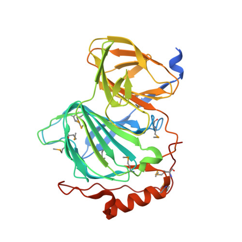
1.78 Angstrom Resolution Crystal Structure of Quercetin 2,3-dioxygenase from Acinetobacter baumannii.
Minasov, G., Shuvalova, L., Brunzelle, J.S., Dubrovska, I., Kiryukhina, O., Endres, M., Anderson, W.F., Satchell, K.J.F., Joachimiak, A.To be published.