6CXX | pdb_00006cxx

Horse liver E267H alcohol dehydrogenase complex with 3'-dephosphocoenzyme A

  • Classification: OXIDOREDUCTASE
  • Organism(s): Equus caballus
  • Expression System: Escherichia coli
  • Mutation(s): Yes 

  • Deposited: 2018-04-04 Released: 2018-04-25 
  • Deposition Author(s): Plapp, B.V.
  • Funding Organization(s): National Institutes of Health/National Human Genome Research Institute (NIH/NHGRI)

Experimental Data Snapshot

  • Method: X-RAY DIFFRACTION
  • Resolution: 1.26 Å
  • R-Value Free: 
    0.208 (Depositor), 0.208 (DCC) 
  • R-Value Work: 
    0.172 (Depositor), 0.172 (DCC) 
  • R-Value Observed: 
    0.173 (Depositor) 

Starting Model: experimental
View more details

wwPDB Validation 3D Report Full Report

Validation slider image for 6CXX

Ligand Structure Quality Assessment 


This is version 1.3 of the entry. See complete history

Literature

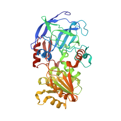
Substitutions of a buried glutamate residue hinder the conformational change in horse liver alcohol dehydrogenase and yield a surprising complex with endogenous 3'-Dephosphocoenzyme A.

Kim, Y.H.Gogerty, D.S.Plapp, B.V.

(2018) Arch Biochem Biophys 653: 97-106

  • DOI: https://doi.org/10.1016/j.abb.2018.07.003
  • Primary Citation Related Structures: 
    6CXX, 6CY3

  • PubMed Abstract: 

    Glu-267 is highly conserved in alcohol dehydrogenases and buried as a negatively-charged residue in a loop of the NAD coenzyme binding domain. Glu-267 might have a structural role and contribute to a rate-promoting vibration that facilitates catalysis. Substitutions of Glu-267 with histidine or asparagine residues increase the dissociation constants for the coenzymes (NAD + by ∼40-fold, NADH by ∼200-fold) and significantly decrease catalytic efficiencies by 16-1200-fold various substrates and substituted enzymes. The turnover numbers modestly change with the substitutions, but hydride transfer is at least partially rate-limiting for turnover for alcohol oxidation. X-ray structures of the E267H and E267 N enzymes are similar to the apoenzyme (open) conformation of the wild-type enzyme, and the substitutions are accommodated by local changes in the structure. Surprisingly, the E267H and E267 N enzymes have endogenous (from the expression in E. coli) 3'-dephosphocoenzyme A bound in the active site with the ADP moiety in the NAD binding site and the pantethiene sulfhydryl bound to the catalytic zinc. The kinetics and crystallography show that the substitutions of Glu-267 hinder the conformational change, which occurs when wild-type enzyme binds coenzymes, and affect productive binding of substrates.


  • Organizational Affiliation
    • Department of Biochemistry, The University of Iowa, Iowa City, IA, 52242, USA. Electronic address: ykim@uwa.edu.

Macromolecule Content 

  • Total Structure Weight: 82.07 kDa 
  • Atom Count: 6,369 
  • Modeled Residue Count: 748 
  • Deposited Residue Count: 748 
  • Unique protein chains: 1

Macromolecules

Find similar proteins by:|  3D Structure
Entity ID: 1
MoleculeChains  Sequence LengthOrganismDetailsImage
Alcohol dehydrogenase E chain
A, B
374Equus caballusMutation(s): 1 
EC: 1.1.1.1
UniProt
Find proteins for P00327 (Equus caballus)
Explore P00327 
Go to UniProtKB:  P00327
Entity Groups
Sequence Clusters30% Identity50% Identity70% Identity90% Identity95% Identity100% Identity
UniProt GroupP00327
Sequence Annotations
Expand
Reference Sequence

Small Molecules

Ligands 4 Unique
IDChains Name / Formula / InChI Key2D Diagram3D Interactions
COD
(Subject of Investigation/LOI)

Query on COD


Download:Ideal Coordinates CCD File
E [auth A],
M [auth B]
DEPHOSPHO COENZYME A
C21 H35 N7 O13 P2 S
KDTSHFARGAKYJN-IBOSZNHHSA-N
MPD

Query on MPD


Download:Ideal Coordinates CCD File
H [auth A](4S)-2-METHYL-2,4-PENTANEDIOL
C6 H14 O2
SVTBMSDMJJWYQN-YFKPBYRVSA-N
MRD

Query on MRD


Download:Ideal Coordinates CCD File
F [auth A],
G [auth A],
I [auth A],
J [auth B],
N [auth B]
(4R)-2-METHYLPENTANE-2,4-DIOL
C6 H14 O2
SVTBMSDMJJWYQN-RXMQYKEDSA-N
ZN
(Subject of Investigation/LOI)

Query on ZN


Download:Ideal Coordinates CCD File
C [auth A],
D [auth A],
K [auth B],
L [auth B]
ZINC ION
Zn
PTFCDOFLOPIGGS-UHFFFAOYSA-N

Experimental Data & Validation

Experimental Data

  • Method: X-RAY DIFFRACTION
  • Resolution: 1.26 Å
  • R-Value Free:  0.208 (Depositor), 0.208 (DCC) 
  • R-Value Work:  0.172 (Depositor), 0.172 (DCC) 
  • R-Value Observed: 0.173 (Depositor) 
Space Group: P 21 21 21
Unit Cell:
Length ( Å )Angle ( ˚ )
a = 55.21α = 90
b = 73.08β = 90
c = 181.61γ = 90
Software Package:
Software NamePurpose
d*TREKdata scaling
REFMACrefinement
PDB_EXTRACTdata extraction
d*TREKdata reduction
AMoREphasing

Structure Validation

View Full Validation Report


Ligand Structure Quality Assessment 


Entry History 

& Funding Information

Deposition Data

  • Released Date: 2018-04-25 
  • Deposition Author(s): Plapp, B.V.

Funding OrganizationLocationGrant Number
National Institutes of Health/National Human Genome Research Institute (NIH/NHGRI)United StatesAA00279

Revision History  (Full details and data files)

  • Version 1.0: 2018-04-25
    Type: Initial release
  • Version 1.1: 2018-08-01
    Changes: Data collection, Database references
  • Version 1.2: 2019-12-18
    Changes: Author supporting evidence
  • Version 1.3: 2023-10-04
    Changes: Data collection, Database references, Derived calculations, Refinement description