6CXK | pdb_00006cxk

E. coli DHFR substrate complex with Dihydrofolate


Experimental Data Snapshot

  • Method: X-RAY DIFFRACTION
  • Resolution: 1.11 Å
  • R-Value Free: 
    0.196 (Depositor) 
  • R-Value Work: 
    0.179 (Depositor) 
  • R-Value Observed: 
    0.179 (Depositor) 

Starting Model: experimental
View more details

wwPDB Validation 3D Report Full Report

Validation slider image for 6CXK

Ligand Structure Quality Assessment 


This is version 1.2 of the entry. See complete history

Literature

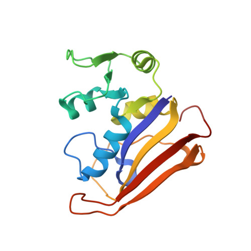
The crystal structure of a tetrahydrofolate-bound dihydrofolate reductase reveals the origin of slow product release.

Cao, H.Gao, M.Zhou, H.Skolnick, J.

(2018) Commun Biol 1: 226-226

  • DOI: https://doi.org/10.1038/s42003-018-0236-y
  • Primary Citation Related Structures: 
    6CQA, 6CW7, 6CXK, 6CYV

  • PubMed Abstract: 

    Dihydrofolate reductase (DHFR) catalyzes the stereospecific reduction of 7,8-dihydrofolate (FH2) to (6s)-5,6,7,8-tetrahydrofolate (FH4) via hydride transfer from NADPH. The consensus Escherichia coli DHFR mechanism involves conformational changes between closed and occluded states occurring during the rate-limiting product release step. Although the Protein Data Bank (PDB) contains over 250 DHFR structures, the FH4 complex structure responsible for rate-limiting product release is unknown. We report to our knowledge the first crystal structure of an E. coli . DHFR:FH4 complex at 1.03 Å resolution showing distinct stabilizing interactions absent in FH2 or related (6R)-5,10-dideaza-FH4 complexes. We discover the time course of decay of the co-purified endogenous FH4 during crystal growth, with conversion from FH4 to FH2 occurring in 2-3 days. We also determine another occluded complex structure of E. coli DHFR with a slow-onset nanomolar inhibitor that contrasts with the methotrexate complex, suggesting a plausible strategy for designing DHFR antibiotics by targeting FH4 product conformations.


  • Organizational Affiliation
    • Center for the Study of Systems Biology, School of Biological Sciences, Georgia Institute of Technology, 950 Atlantic Drive, NW, Atlanta, GA 30332 USA.

Macromolecule Content 

  • Total Structure Weight: 19.52 kDa 
  • Atom Count: 1,637 
  • Modeled Residue Count: 165 
  • Deposited Residue Count: 165 
  • Unique protein chains: 1

Macromolecules

Find similar proteins by:|  3D Structure
Entity ID: 1
MoleculeChains  Sequence LengthOrganismDetailsImage
Dihydrofolate reductase165Escherichia coli CFT073Mutation(s): 0 
Gene Names: folAc0058
EC: 1.5.1.3
UniProt
Find proteins for P0ABQ4 (Escherichia coli (strain K12))
Explore P0ABQ4 
Go to UniProtKB:  P0ABQ4
Entity Groups
Sequence Clusters30% Identity50% Identity70% Identity90% Identity95% Identity100% Identity
UniProt GroupP0ABQ4
Sequence Annotations
Expand
Reference Sequence

Small Molecules

Ligands 3 Unique
IDChains Name / Formula / InChI Key2D Diagram3D Interactions
DHF
(Subject of Investigation/LOI)

Query on DHF


Download:Ideal Coordinates CCD File
K [auth A]DIHYDROFOLIC ACID
C19 H21 N7 O6
OZRNSSUDZOLUSN-LBPRGKRZSA-N
CL

Query on CL


Download:Ideal Coordinates CCD File
J [auth A]CHLORIDE ION
Cl
VEXZGXHMUGYJMC-UHFFFAOYSA-M
MG

Query on MG


Download:Ideal Coordinates CCD File
B [auth A]
C [auth A]
D [auth A]
E [auth A]
F [auth A]
B [auth A],
C [auth A],
D [auth A],
E [auth A],
F [auth A],
G [auth A],
H [auth A],
I [auth A]
MAGNESIUM ION
Mg
JLVVSXFLKOJNIY-UHFFFAOYSA-N

Experimental Data & Validation

Experimental Data

  • Method: X-RAY DIFFRACTION
  • Resolution: 1.11 Å
  • R-Value Free:  0.196 (Depositor) 
  • R-Value Work:  0.179 (Depositor) 
  • R-Value Observed: 0.179 (Depositor) 
Space Group: P 21 21 21
Unit Cell:
Length ( Å )Angle ( ˚ )
a = 33.698α = 90
b = 51.525β = 90
c = 77.421γ = 90
Software Package:
Software NamePurpose
XDSdata reduction
XSCALEdata scaling
PHASERphasing
PHENIXrefinement
PDB_EXTRACTdata extraction

Structure Validation

View Full Validation Report


Ligand Structure Quality Assessment 


Entry History 

& Funding Information

Deposition Data


Funding OrganizationLocationGrant Number
National Institutes of Health/National Institute of General Medical Sciences (NIH/NIGMS)United StatesR35GM118039

Revision History  (Full details and data files)

  • Version 1.0: 2019-01-09
    Type: Initial release
  • Version 1.1: 2020-01-01
    Changes: Author supporting evidence
  • Version 1.2: 2023-10-04
    Changes: Data collection, Database references, Derived calculations, Refinement description