6CXC

3.9A Cryo-EM structure of murine antibody bound at a novel epitope of respiratory syncytial virus fusion protein


Experimental Data Snapshot

  • Method: ELECTRON MICROSCOPY
  • Resolution: 3.90 Å
  • Aggregation State: PARTICLE 
  • Reconstruction Method: SINGLE PARTICLE 

wwPDB Validation   3D Report Full Report


This is version 1.2 of the entry. See complete history


Literature

Structure basis of neutralization by a novel site II/IV antibody against respiratory syncytial virus fusion protein.

Xie, Q.Wang, Z.Ni, F.Chen, X.Ma, J.Patel, N.Lu, H.Liu, Y.Tian, J.H.Flyer, D.Massare, M.J.Ellingsworth, L.Glenn, G.Smith, G.Wang, Q.

(2019) PLoS One 14: e0210749-e0210749

  • DOI: https://doi.org/10.1371/journal.pone.0210749
  • Primary Citation of Related Structures:  
    6CXC

  • PubMed Abstract: 

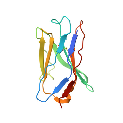
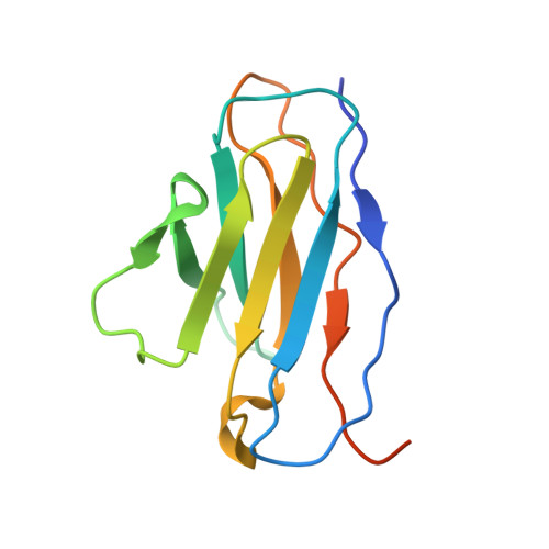
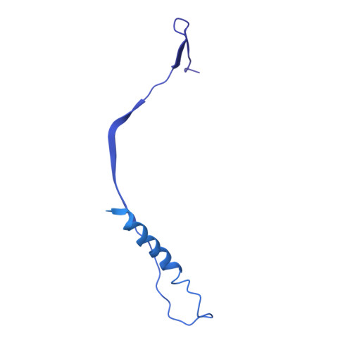
    Globally, human respiratory syncytial virus (RSV) is a leading cause of lower respiratory tract infections in newborns, young children, and the elderly for which there is no vaccine. The RSV fusion (F) glycoprotein is a major target for vaccine development. Here, we describe a novel monoclonal antibody (designated as R4.C6) that recognizes both pre-fusion and post-fusion RSV F, and binds with nanomole affinity to a unique neutralizing site comprised of antigenic sites II and IV on the globular head. A 3.9 Å-resolution structure of RSV F-R4.C6 Fab complex was obtained by single particle cryo-electron microscopy and 3D reconstruction. The structure unraveled detailed interactions of R4.C6 with antigenic site II on one protomer and site IV on a neighboring protomer of post-fusion RSV F protein. These findings significantly further our understanding of the antigenic complexity of the F protein and provide new insights into RSV vaccine design.


  • Organizational Affiliation

    Department of Bioengineering, Rice University, Houston, Texas, United States of America.


Macromolecules
Find similar proteins by:  (by identity cutoff)  |  3D Structure
Entity ID: 1
MoleculeChains Sequence LengthOrganismDetailsImage
R4.C6 Fab Heavy ChainA [auth X],
C [auth G],
E [auth I]
118Mus musculusMutation(s): 0 
Entity Groups  
Sequence Clusters30% Identity50% Identity70% Identity90% Identity95% Identity100% Identity
Sequence Annotations
Expand
  • Reference Sequence
Find similar proteins by:  (by identity cutoff)  |  3D Structure
Entity ID: 2
MoleculeChains Sequence LengthOrganismDetailsImage
R4.C6 Fab Light ChainB [auth Y],
D [auth J],
F [auth K]
122Mus musculusMutation(s): 0 
Entity Groups  
Sequence Clusters30% Identity50% Identity70% Identity90% Identity95% Identity100% Identity
Sequence Annotations
Expand
  • Reference Sequence
Find similar proteins by:  (by identity cutoff)  |  3D Structure
Entity ID: 3
MoleculeChains Sequence LengthOrganismDetailsImage
Fusion glycoprotein F0, Envelope glycoprotein chimera548Human respiratory syncytial virus A2Human immunodeficiency virus 1
This entity is chimeric
Mutation(s): 0 
UniProt
Find proteins for M1E1E4 (Human immunodeficiency virus 1)
Explore M1E1E4 
Go to UniProtKB:  M1E1E4
Find proteins for P03420 (Human respiratory syncytial virus A (strain A2))
Explore P03420 
Go to UniProtKB:  P03420
Entity Groups  
Sequence Clusters30% Identity50% Identity70% Identity90% Identity95% Identity100% Identity
UniProt GroupsM1E1E4P03420
Sequence Annotations
Expand
  • Reference Sequence
Experimental Data & Validation

Experimental Data

  • Method: ELECTRON MICROSCOPY
  • Resolution: 3.90 Å
  • Aggregation State: PARTICLE 
  • Reconstruction Method: SINGLE PARTICLE 
EM Software:
TaskSoftware PackageVersion
RECONSTRUCTIONRELION2.0

Structure Validation

View Full Validation Report


Entry History 

Deposition Data

Revision History  (Full details and data files)

  • Version 1.0: 2019-07-31
    Type: Initial release
  • Version 1.1: 2020-02-12
    Changes: Data collection, Database references
  • Version 1.2: 2020-07-29
    Type: Remediation
    Reason: Carbohydrate remediation
    Changes: Data collection, Derived calculations, Structure summary