6CV7 | pdb_00006cv7

Mouse Protocadherin-15 Extracellular Cadherin Domains 1 through 3


Experimental Data Snapshot

  • Method: X-RAY DIFFRACTION
  • Resolution: 1.69 Å
  • R-Value Free: 
    0.188 (Depositor), 0.199 (DCC) 
  • R-Value Work: 
    0.169 (Depositor), 0.182 (DCC) 
  • R-Value Observed: 
    0.170 (Depositor) 

Starting Model: experimental
View more details

wwPDB Validation 3D Report Full Report

Validation slider image for 6CV7

Ligand Structure Quality Assessment 


This is version 1.4 of the entry. See complete history

Literature

Mechanotransduction by PCDH15 Relies on a Novel cis-Dimeric Architecture.

Dionne, G.Qiu, X.Rapp, M.Liang, X.Zhao, B.Peng, G.Katsamba, P.S.Ahlsen, G.Rubinstein, R.Potter, C.S.Carragher, B.Honig, B.Muller, U.Shapiro, L.

(2018) Neuron 99: 480-492.e5

  • DOI: https://doi.org/10.1016/j.neuron.2018.07.006
  • Primary Citation Related Structures: 
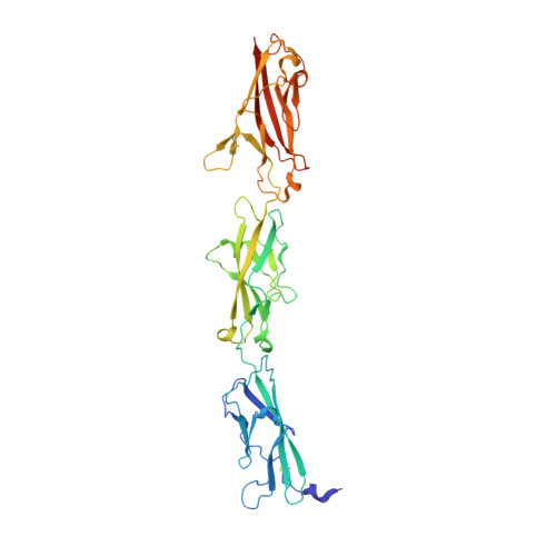
    6CV7

  • PubMed Abstract: 

    The tip link, a filament formed by protocadherin 15 (PCDH15) and cadherin 23, conveys mechanical force from sound waves and head movement to open hair-cell mechanotransduction channels. Tip-link cadherins are thought to have acquired structural features critical for their role in mechanotransduction. Here, we biophysically and structurally characterize the unusual cis-homodimeric architecture of PCDH15. We show that PCDH15 molecules form double-helical assemblies through cis-dimerization interfaces in the extracellular cadherin EC2-EC3 domain region and in a unique membrane-proximal domain. Electron microscopy studies visualize the cis-dimeric PCDH15 assembly and reveal the PCDH15 extracellular domain as a parallel double helix with cis cross-bridges at the two locations we defined. The helical configuration suggests the potential for elasticity through helix winding and unwinding. Functional studies in hair cells show that mutations that perturb PCDH15 dimerization contacts affect mechanotransduction. Together, these data reveal the cis-dimeric architecture of PCDH15 and show that dimerization is critical for sensing mechanical stimuli.


  • Organizational Affiliation
    • Department of Biochemistry and Molecular Biophysics, Columbia University, New York, NY 10032, USA; Zuckerman Mind Brain Behavior Institute, Columbia University, New York, NY 10027, USA.

Macromolecule Content 

  • Total Structure Weight: 44.89 kDa 
  • Atom Count: 3,548 
  • Modeled Residue Count: 365 
  • Deposited Residue Count: 377 
  • Unique protein chains: 1

Macromolecules

Find similar proteins by:|  3D Structure
Entity ID: 1
MoleculeChains  Sequence LengthOrganismDetailsImage
Protocadherin-15377Mus musculusMutation(s): 0 
Gene Names: Pcdh15
UniProt & NIH Common Fund Data Resources
Find proteins for Q99PJ1 (Mus musculus)
Explore Q99PJ1 
Go to UniProtKB:  Q99PJ1
IMPC:  MGI:1891428
Entity Groups
Sequence Clusters30% Identity50% Identity70% Identity90% Identity95% Identity100% Identity
UniProt GroupQ99PJ1
Glycosylation
Glycosylation Sites: 8Go to GlyGen: Q99PJ1-1
Sequence Annotations
Expand
Reference Sequence

Small Molecules

Ligands 4 Unique
IDChains Name / Formula / InChI Key2D Diagram3D Interactions
NAG

Query on NAG


Download:Ideal Coordinates CCD File
Q [auth A],
R [auth A],
S [auth A]
2-acetamido-2-deoxy-beta-D-glucopyranose
C8 H15 N O6
OVRNDRQMDRJTHS-FMDGEEDCSA-N
MAN

Query on MAN


Download:Ideal Coordinates CCD File
L [auth A],
M [auth A],
N [auth A],
O [auth A],
P [auth A]
alpha-D-mannopyranose
C6 H12 O6
WQZGKKKJIJFFOK-PQMKYFCFSA-N
EDO

Query on EDO


Download:Ideal Coordinates CCD File
G [auth A],
H [auth A],
I [auth A],
J [auth A],
K [auth A]
1,2-ETHANEDIOL
C2 H6 O2
LYCAIKOWRPUZTN-UHFFFAOYSA-N
CA

Query on CA


Download:Ideal Coordinates CCD File
B [auth A],
C [auth A],
D [auth A],
E [auth A],
F [auth A]
CALCIUM ION
Ca
BHPQYMZQTOCNFJ-UHFFFAOYSA-N

Experimental Data & Validation

Experimental Data

  • Method: X-RAY DIFFRACTION
  • Resolution: 1.69 Å
  • R-Value Free:  0.188 (Depositor), 0.199 (DCC) 
  • R-Value Work:  0.169 (Depositor), 0.182 (DCC) 
  • R-Value Observed: 0.170 (Depositor) 
Space Group: P 65 2 2
Unit Cell:
Length ( Å )Angle ( ˚ )
a = 114.444α = 90
b = 114.444β = 90
c = 172.011γ = 120
Software Package:
Software NamePurpose
PHENIXrefinement
XDSdata reduction
Aimlessdata scaling
PHASERphasing

Structure Validation

View Full Validation Report


Ligand Structure Quality Assessment 


Entry History 

Deposition Data

Revision History  (Full details and data files)

  • Version 1.0: 2018-08-01
    Type: Initial release
  • Version 1.1: 2020-07-29
    Type: Remediation
    Reason: Carbohydrate remediation
    Changes: Data collection, Derived calculations, Structure summary
  • Version 1.2: 2021-06-23
    Changes: Database references, Structure summary
  • Version 1.3: 2023-10-04
    Changes: Data collection, Database references, Refinement description
  • Version 1.4: 2024-11-20
    Changes: Structure summary