6CUD | pdb_00006cud

Structure of the human TRPC3 in a lipid-occupied, closed state


Experimental Data Snapshot

  • Method: ELECTRON MICROSCOPY
  • Resolution: 3.30 Å
  • Aggregation State: PARTICLE 
  • Reconstruction Method: SINGLE PARTICLE 

wwPDB Validation 3D Report Full Report

Validation slider image for 6CUD

This is version 1.4 of the entry. See complete history

Literature

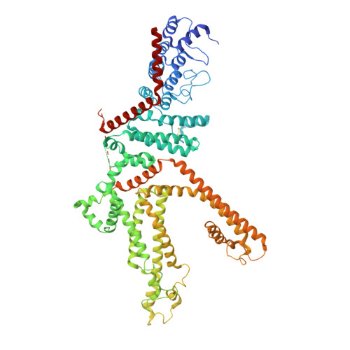
Structure of the human lipid-gated cation channel TRPC3.

Fan, C.Choi, W.Sun, W.Du, J.Lu, W.

(2018) Elife 7

  • DOI: https://doi.org/10.7554/eLife.36852
  • Primary Citation Related Structures: 
    6CUD

  • PubMed Abstract: 

    The TRPC channels are crucially involved in store-operated calcium entry and calcium homeostasis, and they are implicated in human diseases such as neurodegenerative disease, cardiac hypertrophy, and spinocerebellar ataxia. We present a structure of the full-length human TRPC3, a lipid-gated TRPC member, in a lipid-occupied, closed state at 3.3 Angstrom. TRPC3 has four elbow-like membrane reentrant helices prior to the first transmembrane helix. The TRP helix is perpendicular to, and thus disengaged from, the pore-lining S6, suggesting a different gating mechanism from other TRP subfamily channels. The third transmembrane helix S3 is remarkably long, shaping a unique transmembrane domain, and constituting an extracellular domain that may serve as a sensor of external stimuli. We identified two lipid-binding sites, one being sandwiched between the pre-S1 elbow and the S4-S5 linker, and the other being close to the ion-conducting pore, where the conserved LWF motif of the TRPC family is located.


  • Organizational Affiliation
    • Van Andel Institute, Grand Rapids, United States.

Macromolecule Content 

  • Total Structure Weight: 374.45 kDa 
  • Atom Count: 21,028 
  • Modeled Residue Count: 2,816 
  • Deposited Residue Count: 3,224 
  • Unique protein chains: 1

Macromolecules

Find similar proteins by:|  3D Structure
Entity ID: 1
MoleculeChains  Sequence LengthOrganismDetailsImage
Short transient receptor potential channel 3
A, B, C, D
806Homo sapiensMutation(s): 0 
Gene Names: TRPC3TRP3
Membrane Entity: Yes 
UniProt & NIH Common Fund Data Resources
Find proteins for Q13507 (Homo sapiens)
Explore Q13507 
Go to UniProtKB:  Q13507
PHAROS:  Q13507
GTEx:  ENSG00000138741 
Entity Groups
Sequence Clusters30% Identity50% Identity70% Identity90% Identity95% Identity100% Identity
UniProt GroupQ13507
Glycosylation
Glycosylation Sites: 1Go to GlyGen: Q13507-2
Sequence Annotations
Expand
Reference Sequence

Small Molecules

Ligands 3 Unique
IDChains Name / Formula / InChI Key2D Diagram3D Interactions
6OE

Query on 6OE


Download:Ideal Coordinates CCD File
E [auth A],
H [auth B],
K [auth C],
N [auth D]
(2S)-3-{[(S)-(2-aminoethoxy)(hydroxy)phosphoryl]oxy}-2-(hexanoyloxy)propyl hexanoate
C17 H34 N O8 P
PELYUHWUVHDSSU-HNNXBMFYSA-N
FGJ

Query on FGJ


Download:Ideal Coordinates CCD File
F [auth A],
I [auth B],
L [auth C],
O [auth D]
(2R)-3-hydroxypropane-1,2-diyl dihexanoate
C15 H28 O5
DRUFTGMQJWWIOL-CYBMUJFWSA-N
NAG

Query on NAG


Download:Ideal Coordinates CCD File
G [auth A],
J [auth B],
M [auth C],
P [auth D]
2-acetamido-2-deoxy-beta-D-glucopyranose
C8 H15 N O6
OVRNDRQMDRJTHS-FMDGEEDCSA-N

Experimental Data & Validation

Experimental Data

  • Method: ELECTRON MICROSCOPY
  • Resolution: 3.30 Å
  • Aggregation State: PARTICLE 
  • Reconstruction Method: SINGLE PARTICLE 
EM Software:
TaskSoftware PackageVersion
RECONSTRUCTIONFREALIGN9

Structure Validation

View Full Validation Report


Entry History 

Deposition Data

Revision History  (Full details and data files)

  • Version 1.0: 2018-05-16
    Type: Initial release
  • Version 1.1: 2018-05-23
    Changes: Data collection, Database references
  • Version 1.2: 2019-12-18
    Changes: Other
  • Version 1.3: 2020-07-29
    Type: Remediation
    Reason: Carbohydrate remediation
    Changes: Data collection, Derived calculations, Structure summary
  • Version 1.4: 2024-10-30
    Changes: Data collection, Database references, Structure summary