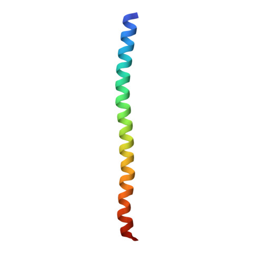
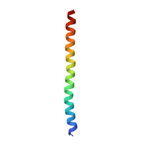
6CSU

The structure of the Cep63-Cep152 heterotetrameric complex


Experimental Data Snapshot

  • Method: X-RAY DIFFRACTION
  • Resolution: 2.50 Å
  • R-Value Free: 0.285 
  • R-Value Work: 0.250 
  • R-Value Observed: 0.254 

wwPDB Validation   3D Report Full Report


This is version 1.1 of the entry. See complete history


Literature

Molecular architecture of a cylindrical self-assembly at human centrosomes.

Kim, T.S.Zhang, L.Il Ahn, J.Meng, L.Chen, Y.Lee, E.Bang, J.K.Lim, J.M.Ghirlando, R.Fan, L.Wang, Y.X.Kim, B.Y.Park, J.E.Lee, K.S.

(2019) Nat Commun 10: 1151-1151

  • DOI: https://doi.org/10.1038/s41467-019-08838-2
  • Primary Citation of Related Structures:  
    6CSU, 6CSV

  • PubMed Abstract: 

    The cell is constructed by higher-order structures and organelles through complex interactions among distinct structural constituents. The centrosome is a membraneless organelle composed of two microtubule-derived structures called centrioles and an amorphous mass of pericentriolar material. Super-resolution microscopic analyses in various organisms revealed that diverse pericentriolar material proteins are concentrically localized around a centriole in a highly organized manner. However, the molecular nature underlying these organizations remains unknown. Here we show that two human pericentriolar material scaffolds, Cep63 and Cep152, cooperatively generate a heterotetrameric α-helical bundle that functions in conjunction with its neighboring hydrophobic motifs to self-assemble into a higher-order cylindrical architecture capable of recruiting downstream components, including Plk4, a key regulator for centriole duplication. Mutations disrupting the self-assembly abrogate Plk4-mediated centriole duplication. Because pericentriolar material organization is evolutionarily conserved, this work may offer a paradigm for investigating the assembly and function of centrosomal scaffolds in various organisms.


  • Organizational Affiliation

    Laboratory of Metabolism, National Cancer Institute, National Institutes of Health, Bethesda, MD, 20892, USA.


Macromolecules
Find similar proteins by:  (by identity cutoff)  |  3D Structure
Entity ID: 1
MoleculeChains Sequence LengthOrganismDetailsImage
Centrosomal protein of 152 kDaA [auth B],
C [auth D]
54Homo sapiensMutation(s): 0 
Gene Names: CEP152KIAA0912
UniProt & NIH Common Fund Data Resources
Find proteins for O94986 (Homo sapiens)
Explore O94986 
Go to UniProtKB:  O94986
PHAROS:  O94986
GTEx:  ENSG00000103995 
Entity Groups  
Sequence Clusters30% Identity50% Identity70% Identity90% Identity95% Identity100% Identity
UniProt GroupO94986
Sequence Annotations
Expand
  • Reference Sequence
Find similar proteins by:  (by identity cutoff)  |  3D Structure
Entity ID: 2
MoleculeChains Sequence LengthOrganismDetailsImage
Centrosomal protein of 63 kDaB [auth C],
D [auth A]
44Homo sapiensMutation(s): 0 
Gene Names: CEP63
UniProt & NIH Common Fund Data Resources
Find proteins for Q96MT8 (Homo sapiens)
Explore Q96MT8 
Go to UniProtKB:  Q96MT8
PHAROS:  Q96MT8
GTEx:  ENSG00000182923 
Entity Groups  
Sequence Clusters30% Identity50% Identity70% Identity90% Identity95% Identity100% Identity
UniProt GroupQ96MT8
Sequence Annotations
Expand
  • Reference Sequence
Experimental Data & Validation

Experimental Data

  • Method: X-RAY DIFFRACTION
  • Resolution: 2.50 Å
  • R-Value Free: 0.285 
  • R-Value Work: 0.250 
  • R-Value Observed: 0.254 
  • Space Group: P 21 21 21
Unit Cell:
Length ( Å )Angle ( ˚ )
a = 28.066α = 90
b = 41.523β = 90
c = 205.619γ = 90
Software Package:
Software NamePurpose
PHENIXrefinement
HKL-2000data collection
HKL-2000data reduction
HKL-2000data scaling
PHENIXphasing
Cootmodel building

Structure Validation

View Full Validation Report


Entry History 

Deposition Data

Revision History  (Full details and data files)

  • Version 1.0: 2019-03-27
    Type: Initial release
  • Version 1.1: 2024-03-13
    Changes: Data collection, Database references