6CSS

Crystal structure of Danio rerio histone deacetylase 6 catalytic domain 2 in complex with cyclopentenylhydroxamate


Experimental Data Snapshot

  • Method: X-RAY DIFFRACTION
  • Resolution: 1.70 Å
  • R-Value Free: 0.193 
  • R-Value Work: 0.170 
  • R-Value Observed: 0.171 

wwPDB Validation   3D Report Full Report


Ligand Structure Quality Assessment 


This is version 1.3 of the entry. See complete history


Literature

Entropy as a Driver of Selectivity for Inhibitor Binding to Histone Deacetylase 6.

Porter, N.J.Wagner, F.F.Christianson, D.W.

(2018) Biochemistry 57: 3916-3924

  • DOI: https://doi.org/10.1021/acs.biochem.8b00367
  • Primary Citation of Related Structures:  
    6CSP, 6CSQ, 6CSR, 6CSS

  • PubMed Abstract: 

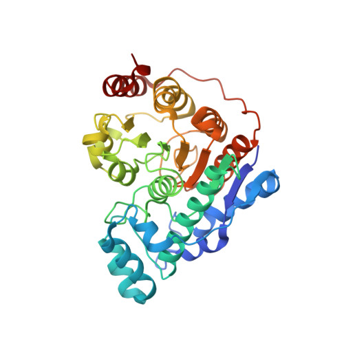
    Among the metal-dependent histone deacetylases, the class IIb isozyme HDAC6 is remarkable because of its role in the regulation of microtubule dynamics in the cytosol. Selective inhibition of HDAC6 results in microtubule hyperacetylation, leading to cell cycle arrest and apoptosis, which is a validated strategy for cancer chemotherapy and the treatment of other disorders. HDAC6 inhibitors generally consist of a Zn 2+ -binding group such as a hydroxamate, a linker, and a capping group; the capping group is a critical determinant of isozyme selectivity. Surprisingly, however, even "capless" inhibitors exhibit appreciable HDAC6 selectivity. To probe the chemical basis for this selectivity, we now report high-resolution crystal structures of HDAC6 complexed with capless cycloalkyl hydroxamate inhibitors 1-4. Each inhibitor hydroxamate group coordinates to the catalytic Zn 2+ ion with canonical bidentate geometry. Additionally, the olefin moieties of compounds 2 and 4 bind in an aromatic crevice between the side chains of F583 and F643. Reasoning that similar binding could be achieved in the representative class I isozyme HDAC8, we employed isothermal titration calorimetry to study the thermodynamics of inhibitor binding. These measurements indicate that the entropy of inhibitor binding is generally positive for binding to HDAC6 and negative for binding to HDAC8, resulting in ≤313-fold selectivity for binding to HDAC6 relative to HDAC8. Thus, favorable binding entropy contributes to HDAC6 selectivity. Notably, cyclohexenyl hydroxamate 2 represents a promising lead for derivatization with capping groups that may further enhance its impressive 313-fold thermodynamic selectivity for HDAC6 inhibition.


  • Organizational Affiliation

    Roy and Diana Vagelos Laboratories, Department of Chemistry , University of Pennsylvania , 231 South 34th Street , Philadelphia , Pennsylvania 19104-6323 , United States.


Macromolecules
Find similar proteins by:  (by identity cutoff)  |  3D Structure
Entity ID: 1
MoleculeChains Sequence LengthOrganismDetailsImage
Hdac6 protein
A, B
364Danio rerioMutation(s): 0 
Gene Names: hdac6
UniProt
Find proteins for F8W4B7 (Danio rerio)
Explore F8W4B7 
Go to UniProtKB:  F8W4B7
Entity Groups  
Sequence Clusters30% Identity50% Identity70% Identity90% Identity95% Identity100% Identity
UniProt GroupF8W4B7
Sequence Annotations
Expand
  • Reference Sequence
Small Molecules
Ligands 4 Unique
IDChains Name / Formula / InChI Key2D Diagram3D Interactions
FBJ (Subject of Investigation/LOI)
Query on FBJ

Download Ideal Coordinates CCD File 
F [auth A],
P [auth B]
N-hydroxycyclopent-1-ene-1-carboxamide
C6 H9 N O2
RYYGSXXWQSXKRP-UHFFFAOYSA-N
ZN
Query on ZN

Download Ideal Coordinates CCD File 
C [auth A],
M [auth B]
ZINC ION
Zn
PTFCDOFLOPIGGS-UHFFFAOYSA-N
EDO
Query on EDO

Download Ideal Coordinates CCD File 
G [auth A]
H [auth A]
I [auth A]
J [auth A]
K [auth A]
G [auth A],
H [auth A],
I [auth A],
J [auth A],
K [auth A],
L [auth A],
Q [auth B],
R [auth B],
S [auth B]
1,2-ETHANEDIOL
C2 H6 O2
LYCAIKOWRPUZTN-UHFFFAOYSA-N
K
Query on K

Download Ideal Coordinates CCD File 
D [auth A],
E [auth A],
N [auth B],
O [auth B]
POTASSIUM ION
K
NPYPAHLBTDXSSS-UHFFFAOYSA-N
Binding Affinity Annotations 
IDSourceBinding Affinity
FBJ Binding MOAD:  6CSS Kd: 25 (nM) from 1 assay(s)
Experimental Data & Validation

Experimental Data

  • Method: X-RAY DIFFRACTION
  • Resolution: 1.70 Å
  • R-Value Free: 0.193 
  • R-Value Work: 0.170 
  • R-Value Observed: 0.171 
  • Space Group: P 21 21 21
Unit Cell:
Length ( Å )Angle ( ˚ )
a = 74.791α = 90
b = 92.021β = 90
c = 96.64γ = 90
Software Package:
Software NamePurpose
MOSFLMdata reduction
Aimlessdata scaling
PHASERphasing
PHENIXrefinement
PDB_EXTRACTdata extraction

Structure Validation

View Full Validation Report


Ligand Structure Quality Assessment 


Entry History & Funding Information

Deposition Data


Funding OrganizationLocationGrant Number
National Institutes of Health/National Institute of General Medical Sciences (NIH/NIGMS)United StatesGM49758

Revision History  (Full details and data files)

  • Version 1.0: 2018-05-30
    Type: Initial release
  • Version 1.1: 2018-07-11
    Changes: Data collection, Database references
  • Version 1.2: 2020-01-01
    Changes: Author supporting evidence
  • Version 1.3: 2024-03-13
    Changes: Data collection, Database references, Derived calculations