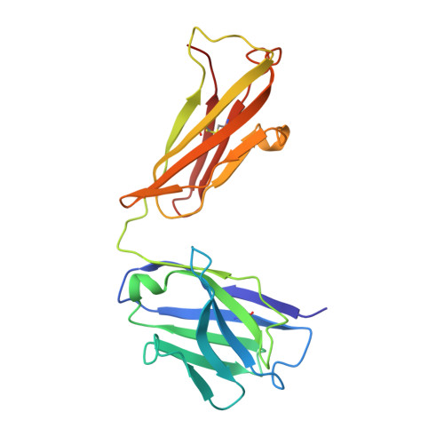
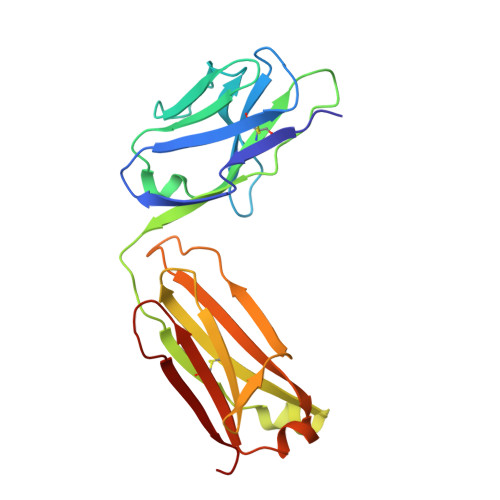
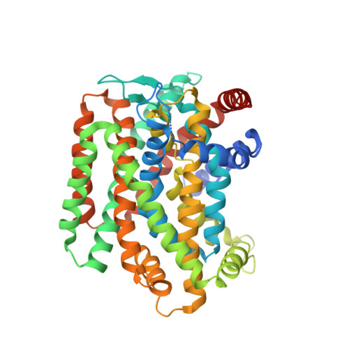
6CSE

Crystal structure of sodium/alanine symporter AgcS with L-alanine bound


Experimental Data Snapshot

  • Method: X-RAY DIFFRACTION
  • Resolution: 3.24 Å
  • R-Value Free: 0.251 
  • R-Value Work: 0.230 
  • R-Value Observed: 0.231 

wwPDB Validation   3D Report Full Report


This is version 1.1 of the entry. See complete history


Literature

Structural basis for substrate binding and specificity of a sodium-alanine symporter AgcS.

Ma, J.Lei, H.T.Reyes, F.E.Sanchez-Martinez, S.Sarhan, M.F.Hattne, J.Gonen, T.

(2019) Proc Natl Acad Sci U S A 116: 2086-2090

  • DOI: https://doi.org/10.1073/pnas.1806206116
  • Primary Citation of Related Structures:  
    6CSE, 6CSF

  • PubMed Abstract: 

    The amino acid, polyamine, and organocation (APC) superfamily is the second largest superfamily of membrane proteins forming secondary transporters that move a range of organic molecules across the cell membrane. Each transporter in the APC superfamily is specific for a unique subset of substrates, even if they possess a similar structural fold. The mechanism of substrate selectivity remains, by and large, elusive. Here, we report two crystal structures of an APC member from Methanococcus maripaludis , the alanine or glycine:cation symporter (AgcS), with l- or d-alanine bound. Structural analysis combined with site-directed mutagenesis and functional studies inform on substrate binding, specificity, and modulation of the AgcS family and reveal key structural features that allow this transporter to accommodate glycine and alanine while excluding all other amino acids. Mutation of key residues in the substrate binding site expand the selectivity to include valine and leucine. These studies provide initial insights into substrate selectivity in AgcS symporters.


  • Organizational Affiliation

    Janelia Research Campus, Howard Hughes Medical Institute, Ashburn, VA 20147.


Macromolecules
Find similar proteins by:  (by identity cutoff)  |  3D Structure
Entity ID: 1
MoleculeChains Sequence LengthOrganismDetailsImage
Monoclonal antibody FAB heavy chainA,
D [auth H]
214Mus musculusMutation(s): 0 
Entity Groups  
Sequence Clusters30% Identity50% Identity70% Identity90% Identity95% Identity100% Identity
Sequence Annotations
Expand
  • Reference Sequence
Find similar proteins by:  (by identity cutoff)  |  3D Structure
Entity ID: 2
MoleculeChains Sequence LengthOrganismDetailsImage
Monoclonal antibody FAB light chainB,
F [auth L]
213Mus musculusMutation(s): 0 
Entity Groups  
Sequence Clusters30% Identity50% Identity70% Identity90% Identity95% Identity100% Identity
Sequence Annotations
Expand
  • Reference Sequence
Find similar proteins by:  (by identity cutoff)  |  3D Structure
Entity ID: 3
MoleculeChains Sequence LengthOrganismDetailsImage
Sodium/alanine symporter AgcSC [auth M],
E [auth C]
453Methanococcus maripaludis S2Mutation(s): 0 
Gene Names: agcSMMP1511
Membrane Entity: Yes 
UniProt
Find proteins for Q6LX42 (Methanococcus maripaludis (strain DSM 14266 / JCM 13030 / NBRC 101832 / S2 / LL))
Explore Q6LX42 
Go to UniProtKB:  Q6LX42
Entity Groups  
Sequence Clusters30% Identity50% Identity70% Identity90% Identity95% Identity100% Identity
UniProt GroupQ6LX42
Sequence Annotations
Expand
  • Reference Sequence
Small Molecules
Ligands 2 Unique
IDChains Name / Formula / InChI Key2D Diagram3D Interactions
ALA
Query on ALA

Download Ideal Coordinates CCD File 
H [auth M]ALANINE
C3 H7 N O2
QNAYBMKLOCPYGJ-REOHCLBHSA-N
NA
Query on NA

Download Ideal Coordinates CCD File 
G [auth M]SODIUM ION
Na
FKNQFGJONOIPTF-UHFFFAOYSA-N
Experimental Data & Validation

Experimental Data

  • Method: X-RAY DIFFRACTION
  • Resolution: 3.24 Å
  • R-Value Free: 0.251 
  • R-Value Work: 0.230 
  • R-Value Observed: 0.231 
  • Space Group: P 61
Unit Cell:
Length ( Å )Angle ( ˚ )
a = 183.477α = 90
b = 183.477β = 90
c = 349.067γ = 120
Software Package:
Software NamePurpose
PHENIXrefinement
XSCALEdata scaling
XDSdata reduction
PHASERphasing
PDB_EXTRACTdata extraction

Structure Validation

View Full Validation Report


Entry History 

Deposition Data

Revision History  (Full details and data files)

  • Version 1.0: 2019-01-30
    Type: Initial release
  • Version 1.1: 2019-02-20
    Changes: Data collection, Database references