6CO1 | pdb_00006co1

Structure of human TIRR in complex with 53BP1 Tudor domains

  • Classification: PROTEIN BINDING
  • Organism(s): Homo sapiens
  • Expression System: Escherichia coli
  • Mutation(s): No 

  • Deposited: 2018-03-10 Released: 2018-06-06 
  • Deposition Author(s): Cui, G., Botuyan, M.V., Mer, G.
  • Funding Organization(s): National Institutes of Health/National Cancer Institute (NIH/NCI), National Institutes of Health/National Institute of General Medical Sciences (NIH/NIGMS), Department of Defense Ovarian Cancer Research

Experimental Data Snapshot

  • Method: X-RAY DIFFRACTION
  • Resolution: 2.18 Å
  • R-Value Free: 
    0.203 (Depositor), 0.197 (DCC) 
  • R-Value Work: 
    0.164 (Depositor), 0.160 (DCC) 
  • R-Value Observed: 
    0.167 (Depositor) 

Starting Models: experimental
View more details

wwPDB Validation 3D Report Full Report

Validation slider image for 6CO1

This is version 1.4 of the entry. See complete history

Literature

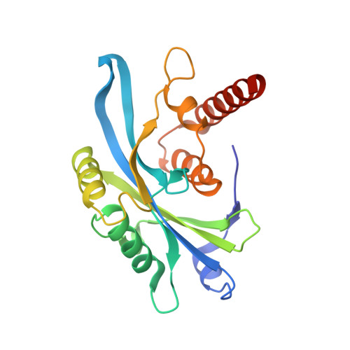
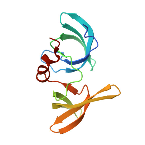
Mechanism of 53BP1 activity regulation by RNA-binding TIRR and a designer protein.

Botuyan, M.V.Cui, G.Drane, P.Oliveira, C.Detappe, A.Brault, M.E.Parnandi, N.Chaubey, S.Thompson, J.R.Bragantini, B.Zhao, D.Chapman, J.R.Chowdhury, D.Mer, G.

(2018) Nat Struct Mol Biol 25: 591-600

  • DOI: https://doi.org/10.1038/s41594-018-0083-z
  • Primary Citation Related Structures: 
    6CO1, 6CO2, 6D0L

  • PubMed Abstract: 

    Dynamic protein interaction networks such as DNA double-strand break (DSB) signaling are modulated by post-translational modifications. The DNA repair factor 53BP1 is a rare example of a protein whose post-translational modification-binding function can be switched on and off. 53BP1 is recruited to DSBs by recognizing histone lysine methylation within chromatin, an activity directly inhibited by the 53BP1-binding protein TIRR. X-ray crystal structures of TIRR and a designer protein bound to 53BP1 now reveal a unique regulatory mechanism in which an intricate binding area centered on an essential TIRR arginine residue blocks the methylated-chromatin-binding surface of 53BP1. A 53BP1 separation-of-function mutation that abolishes TIRR-mediated regulation in cells renders 53BP1 hyperactive in response to DSBs, highlighting the key inhibitory function of TIRR. This 53BP1 inhibition is relieved by TIRR-interacting RNA molecules, providing proof-of-principle of RNA-triggered 53BP1 recruitment to DSBs.


  • Organizational Affiliation
    • Department of Biochemistry and Molecular Biology, Mayo Clinic, Rochester, MN, USA.

Macromolecule Content 

  • Total Structure Weight: 120.05 kDa 
  • Atom Count: 9,484 
  • Modeled Residue Count: 1,043 
  • Deposited Residue Count: 1,074 
  • Unique protein chains: 2

Macromolecules

Find similar proteins by:|  3D Structure
Entity ID: 1
MoleculeChains  Sequence LengthOrganismDetailsImage
Tudor-interacting repair regulator protein
A, B, C, D
207Homo sapiensMutation(s): 0 
Gene Names: NUDT16L1SDOSTIRR
UniProt & NIH Common Fund Data Resources
Find proteins for Q9BRJ7 (Homo sapiens)
Explore Q9BRJ7 
Go to UniProtKB:  Q9BRJ7
GTEx:  ENSG00000168101 
Entity Groups
Sequence Clusters30% Identity50% Identity70% Identity90% Identity95% Identity100% Identity
UniProt GroupQ9BRJ7
Sequence Annotations
Expand
Reference Sequence
Find similar proteins by:|  3D Structure
Entity ID: 2
MoleculeChains  Sequence LengthOrganismDetailsImage
TP53-binding protein 1
E, F
123Homo sapiensMutation(s): 0 
Gene Names: TP53BP1
UniProt & NIH Common Fund Data Resources
Find proteins for Q12888 (Homo sapiens)
Explore Q12888 
Go to UniProtKB:  Q12888
PHAROS:  Q12888
GTEx:  ENSG00000067369 
Entity Groups
Sequence Clusters30% Identity50% Identity70% Identity90% Identity95% Identity100% Identity
UniProt GroupQ12888
Sequence Annotations
Expand
Reference Sequence

Experimental Data & Validation

Experimental Data

  • Method: X-RAY DIFFRACTION
  • Resolution: 2.18 Å
  • R-Value Free:  0.203 (Depositor), 0.197 (DCC) 
  • R-Value Work:  0.164 (Depositor), 0.160 (DCC) 
  • R-Value Observed: 0.167 (Depositor) 
Space Group: P 1
Unit Cell:
Length ( Å )Angle ( ˚ )
a = 61.84α = 85.24
b = 77.03β = 86.07
c = 77.24γ = 86.09
Software Package:
Software NamePurpose
PHENIXrefinement
HKL-2000data reduction
HKL-2000data scaling
PHENIXphasing

Structure Validation

View Full Validation Report


Entry History 

& Funding Information

Deposition Data


Funding OrganizationLocationGrant Number
National Institutes of Health/National Cancer Institute (NIH/NCI)United StatesCA132878
National Institutes of Health/National Institute of General Medical Sciences (NIH/NIGMS)United StatesGM116829
Department of Defense Ovarian Cancer ResearchUnited StatesW81XWH-16-1-0391

Revision History  (Full details and data files)

  • Version 1.0: 2018-06-06
    Type: Initial release
  • Version 1.1: 2018-07-11
    Changes: Data collection, Database references
  • Version 1.2: 2018-07-25
    Changes: Data collection, Database references
  • Version 1.3: 2019-12-04
    Changes: Author supporting evidence
  • Version 1.4: 2023-10-04
    Changes: Advisory, Data collection, Database references, Refinement description