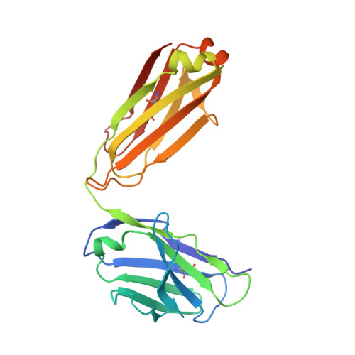
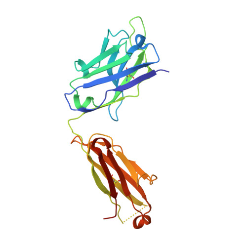
6CNR

aducanumab apo Fab


Experimental Data Snapshot

  • Method: X-RAY DIFFRACTION
  • Resolution: 2.09 Å
  • R-Value Free: 0.239 
  • R-Value Work: 0.211 
  • R-Value Observed: 0.212 

wwPDB Validation   3D Report Full Report


This is version 1.2 of the entry. See complete history


Literature

Structural and kinetic basis for the selectivity of aducanumab for aggregated forms of amyloid-beta.

Arndt, J.W.Qian, F.Smith, B.A.Quan, C.Kilambi, K.P.Bush, M.W.Walz, T.Pepinsky, R.B.Bussiere, T.Hamann, S.Cameron, T.O.Weinreb, P.H.

(2018) Sci Rep 8: 6412-6412

  • DOI: https://doi.org/10.1038/s41598-018-24501-0
  • Primary Citation of Related Structures:  
    6CNR, 6CO3

  • PubMed Abstract: 

    Aducanumab, a human-derived antibody targeting amyloid-β (Aβ), is in Phase 3 clinical trials for the treatment of Alzheimer's disease. Biochemical and structural analyses show that aducanumab binds a linear epitope formed by amino acids 3-7 of the Aβ peptide. Aducanumab discriminates between monomers and oligomeric or fibrillar aggregates based on weak monovalent affinity, fast binding kinetics and strong avidity for epitope-rich aggregates. Direct comparative studies with analogs of gantenerumab, bapineuzumab and solanezumab demonstrate clear differentiation in the binding properties of these antibodies. The crystal structure of the Fab fragment of aducanumab bound to its epitope peptide reveals that aducanumab binds to the N terminus of Aβ in an extended conformation, distinct from those seen in structures with other antibodies that target this immunodominant epitope. Aducanumab recognizes a compact epitope that sits in a shallow pocket on the antibody surface. In silico analyses suggest that aducanumab interacts weakly with the Aβ monomer and may accommodate a variety of peptide conformations, further supporting its selectivity for Aβ aggregates. Our studies provide a structural rationale for the low affinity of aducanumab for non-pathogenic monomers and its greater selectivity for aggregated forms than is seen for other Aβ-targeting antibodies.


  • Organizational Affiliation

    Biogen, Cambridge, MA, USA. joe.arndt@biogen.com.


Macromolecules
Find similar proteins by:  (by identity cutoff)  |  3D Structure
Entity ID: 1
MoleculeChains Sequence LengthOrganismDetailsImage
Aducanumab light chainA [auth L]214Homo sapiensMutation(s): 0 
Entity Groups  
Sequence Clusters30% Identity50% Identity70% Identity90% Identity95% Identity100% Identity
Sequence Annotations
Expand
  • Reference Sequence
Find similar proteins by:  (by identity cutoff)  |  3D Structure
Entity ID: 2
MoleculeChains Sequence LengthOrganismDetailsImage
Aducanumab heavy chainB [auth H]227Homo sapiensMutation(s): 0 
Entity Groups  
Sequence Clusters30% Identity50% Identity70% Identity90% Identity95% Identity100% Identity
Sequence Annotations
Expand
  • Reference Sequence
Small Molecules
Ligands 1 Unique
IDChains Name / Formula / InChI Key2D Diagram3D Interactions
SO4
Query on SO4

Download Ideal Coordinates CCD File 
C [auth H]SULFATE ION
O4 S
QAOWNCQODCNURD-UHFFFAOYSA-L
Experimental Data & Validation

Experimental Data

  • Method: X-RAY DIFFRACTION
  • Resolution: 2.09 Å
  • R-Value Free: 0.239 
  • R-Value Work: 0.211 
  • R-Value Observed: 0.212 
  • Space Group: C 1 2 1
Unit Cell:
Length ( Å )Angle ( ˚ )
a = 143.311α = 90
b = 64.126β = 95.85
c = 67.293γ = 90
Software Package:
Software NamePurpose
PHENIXrefinement
HKL-2000data reduction
SCALEPACKdata scaling
MOLREPphasing

Structure Validation

View Full Validation Report


Entry History 

Deposition Data

  • Released Date: 2018-05-02 
  • Deposition Author(s): Arndt, J.W.

Revision History  (Full details and data files)

  • Version 1.0: 2018-05-02
    Type: Initial release
  • Version 1.1: 2018-05-09
    Changes: Data collection, Database references
  • Version 1.2: 2023-10-04
    Changes: Data collection, Database references, Refinement description