6CNL | pdb_00006cnl

Crystal Structure of H105A PGAM5 Dodecamer


Experimental Data Snapshot

  • Method: X-RAY DIFFRACTION
  • Resolution: 2.60 Å
  • R-Value Free: 
    0.264 (Depositor), 0.261 (DCC) 
  • R-Value Work: 
    0.223 (Depositor), 0.225 (DCC) 
  • R-Value Observed: 
    0.225 (Depositor) 

Starting Model: experimental
View more details

wwPDB Validation 3D Report Full Report

Validation slider image for 6CNL

This is version 1.1 of the entry. See complete history

Literature

Functional role of PGAM5 multimeric assemblies and their polymerization into filaments.

Ruiz, K.Thaker, T.M.Agnew, C.Miller-Vedam, L.Trenker, R.Herrera, C.Ingaramo, M.Toso, D.Frost, A.Jura, N.

(2019) Nat Commun 10: 531-531

  • DOI: https://doi.org/10.1038/s41467-019-08393-w
  • Primary Citation Related Structures: 
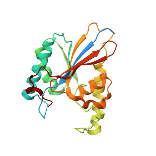
    6CNI, 6CNL

  • PubMed Abstract: 

    PGAM5 is a mitochondrial protein phosphatase whose genetic ablation in mice results in mitochondria-related disorders, including neurodegeneration. Functions of PGAM5 include regulation of mitophagy, cell death, metabolism and aging. However, mechanisms regulating PGAM5 activation and signaling are poorly understood. Using electron cryo-microscopy, we show that PGAM5 forms dodecamers in solution. We also present a crystal structure of PGAM5 that reveals the determinants of dodecamer formation. Furthermore, we observe PGAM5 dodecamer assembly into filaments both in vitro and in cells. We find that PGAM5 oligomerization into a dodecamer is not only essential for catalytic activation, but this form also plays a structural role on mitochondrial membranes, which is independent of phosphatase activity. Together, these findings suggest that modulation of the oligomerization of PGAM5 may be a regulatory switch of potential therapeutic interest.


  • Organizational Affiliation
    • Cardiovascular Research Institute, University of California San Francisco, San Francisco, CA, 94158, USA.

Macromolecule Content 

  • Total Structure Weight: 325.84 kDa 
  • Atom Count: 18,873 
  • Modeled Residue Count: 2,379 
  • Deposited Residue Count: 2,844 
  • Unique protein chains: 2

Macromolecules

Find similar proteins by:|  3D Structure
Entity ID: 1
MoleculeChains  Sequence LengthOrganismDetailsImage
Serine/threonine-protein phosphatase PGAM5, mitochondrial223Homo sapiensMutation(s): 1 
Gene Names: PGAM5
EC: 3.1.3.16
UniProt & NIH Common Fund Data Resources
Find proteins for Q96HS1 (Homo sapiens)
Explore Q96HS1 
Go to UniProtKB:  Q96HS1
PHAROS:  Q96HS1
GTEx:  ENSG00000247077 
Entity Groups
Sequence Clusters30% Identity50% Identity70% Identity90% Identity95% Identity100% Identity
UniProt GroupQ96HS1
Sequence Annotations
Expand
Reference Sequence
Find similar proteins by:  Sequence   |   3D Structure  
Entity ID: 2
MoleculeChains  Sequence LengthOrganismDetailsImage
PGAM5 Multimerization Motif Peptide14Homo sapiensMutation(s): 0 

Small Molecules

Ligands 1 Unique
IDChains Name / Formula / InChI Key2D Diagram3D Interactions
MG

Query on MG


Download:Ideal Coordinates CCD File
AA [auth C]
AB [auth E]
BA [auth C]
BB [auth E]
CA [auth C]
AA [auth C],
AB [auth E],
BA [auth C],
BB [auth E],
CA [auth C],
CB [auth P],
DA [auth C],
EA [auth L],
FA [auth L],
GA [auth J],
HA [auth J],
IA [auth F],
JA [auth F],
KA [auth F],
LA [auth F],
MA [auth H],
NA [auth H],
OA [auth K],
PA [auth D],
QA [auth D],
RA [auth D],
SA [auth B],
TA [auth B],
UA [auth I],
VA [auth I],
WA [auth I],
XA [auth G],
Y [auth A],
YA [auth G],
Z [auth C],
ZA [auth G]
MAGNESIUM ION
Mg
JLVVSXFLKOJNIY-UHFFFAOYSA-N

Experimental Data & Validation

Experimental Data

  • Method: X-RAY DIFFRACTION
  • Resolution: 2.60 Å
  • R-Value Free:  0.264 (Depositor), 0.261 (DCC) 
  • R-Value Work:  0.223 (Depositor), 0.225 (DCC) 
  • R-Value Observed: 0.225 (Depositor) 
Space Group: P 21 21 21
Unit Cell:
Length ( Å )Angle ( ˚ )
a = 49.372α = 90
b = 242.538β = 90
c = 272.458γ = 90
Software Package:
Software NamePurpose
PHENIXrefinement
XDSdata reduction
Aimlessdata scaling
PHASERphasing

Structure Validation

View Full Validation Report


Entry History 

Deposition Data

Revision History  (Full details and data files)

  • Version 1.0: 2019-02-13
    Type: Initial release
  • Version 1.1: 2023-10-04
    Changes: Data collection, Database references, Derived calculations, Refinement description