6CHE

Selenomethionine mutant (A34Sem) of protein GB1 examined by X-ray diffraction

  • Classification: IMMUNE SYSTEM
  • Organism(s): Streptococcus sp. 'group G
  • Expression System: Escherichia coli BL21(DE3)
  • Mutation(s): Yes 

  • Deposited: 2018-02-22 Released: 2019-07-10 
  • Deposition Author(s): Chen, Q.
  • Funding Organization(s): National Institutes of Health/National Institute of General Medical Sciences (NIH/NIGMS), National Science Foundation (NSF, United States)

Experimental Data Snapshot

  • Method: X-RAY DIFFRACTION
  • Resolution: 1.10 Å
  • R-Value Free: 0.147 
  • R-Value Work: 0.129 
  • R-Value Observed: 0.131 

wwPDB Validation   3D Report Full Report


This is version 1.5 of the entry. See complete history


Literature

77Se NMR Probes the Protein Environment of Selenomethionine.

Chen, Q.Xu, S.Lu, X.Boeri, M.V.Pepelyayeva, Y.Diaz, E.L.Soni, S.D.Allaire, M.Forstner, M.B.Bahnson, B.J.Rozovsky, S.

(2020) J Phys Chem B 124: 601-616

  • DOI: https://doi.org/10.1021/acs.jpcb.9b07466
  • Primary Citation of Related Structures:  
    6C9O, 6CHE, 6CNE, 6CPZ, 6CTE

  • PubMed Abstract: 

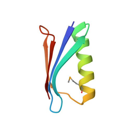
    Sulfur is critical for the correct structure and proper function of proteins. Yet, lacking a sensitive enough isotope, nuclear magnetic resonance (NMR) experiments are unable to deliver for sulfur in proteins the usual wealth of chemical, dynamic, and structural information. This limitation can be circumvented by substituting sulfur with selenium, which has similar physicochemical properties and minimal impact on protein structures but possesses an NMR compatible isotope ( 77 Se). Here we exploit the sensitivity of 77 Se NMR to the nucleus' chemical milieu and use selenomethionine as a probe for its proteinaceous environment. However, such selenium NMR spectra of proteins currently resist a reliable interpretation because systematic connections between variations of system variables and changes in 77 Se NMR parameters are still lacking. To start narrowing this knowledge gap, we report here on a biological 77 Se magnetic resonance data bank based on a systematically designed library of GB1 variants in which a single selenomethionine was introduced at different locations within the protein. We recorded the resulting isotropic 77 Se chemical shifts and relaxation times for six GB1 variants by solution-state 77 Se NMR. For four of the GB1 variants we were also able to determine the chemical shift anisotropy tensor of SeM by solid-state 77 Se NMR. To enable interpretation of the NMR data, the structures of five of the GB1 variants were solved by X-ray crystallography to a resolution of 1.2 Å, allowing us to unambiguously determine the conformation of the selenomethionine. Finally, we combine our solution- and solid-state NMR data with the structural information to arrive at general insights regarding the execution and interpretation of 77 Se NMR experiments that exploit selenomethionine to probe proteins.


  • Organizational Affiliation

    Department of Chemistry and Biochemistry , University of Delaware , Newark , Delaware 19716 , United States.


Macromolecules
Find similar proteins by:  (by identity cutoff)  |  3D Structure
Entity ID: 1
MoleculeChains Sequence LengthOrganismDetailsImage
Immunoglobulin G-binding protein G56Streptococcus sp. 'group GMutation(s): 1 
Gene Names: spg
UniProt
Find proteins for P19909 (Streptococcus sp. group G)
Explore P19909 
Go to UniProtKB:  P19909
Entity Groups  
Sequence Clusters30% Identity50% Identity70% Identity90% Identity95% Identity100% Identity
UniProt GroupP19909
Sequence Annotations
Expand
  • Reference Sequence
Small Molecules
Modified Residues  1 Unique
IDChains TypeFormula2D DiagramParent
MSE
Query on MSE
A
L-PEPTIDE LINKINGC5 H11 N O2 SeMET
Experimental Data & Validation

Experimental Data

  • Method: X-RAY DIFFRACTION
  • Resolution: 1.10 Å
  • R-Value Free: 0.147 
  • R-Value Work: 0.129 
  • R-Value Observed: 0.131 
  • Space Group: P 1 21 1
Unit Cell:
Length ( Å )Angle ( ˚ )
a = 22.965α = 90
b = 37.048β = 110.16
c = 28.719γ = 90
Software Package:
Software NamePurpose
PHENIXrefinement
XDSdata reduction
PHENIXmodel building

Structure Validation

View Full Validation Report


Entry History & Funding Information

Deposition Data

  • Released Date: 2019-07-10 
  • Deposition Author(s): Chen, Q.

Funding OrganizationLocationGrant Number
National Institutes of Health/National Institute of General Medical Sciences (NIH/NIGMS)United States5 P30 GM110758-02
National Institutes of Health/National Institute of General Medical Sciences (NIH/NIGMS)United States5 P20 GM104316
National Science Foundation (NSF, United States)United StatesMCB-1616178

Revision History  (Full details and data files)

  • Version 1.0: 2019-07-10
    Type: Initial release
  • Version 1.1: 2019-11-27
    Changes: Author supporting evidence
  • Version 1.2: 2020-01-22
    Changes: Database references
  • Version 1.3: 2020-02-12
    Changes: Database references
  • Version 1.4: 2023-10-04
    Changes: Data collection, Database references, Refinement description
  • Version 1.5: 2023-11-15
    Changes: Data collection