An endogenous dAMP ligand inBacillus subtilisclass Ib RNR promotes assembly of a noncanonical dimer for regulation by dATP.
Parker, M.J., Maggiolo, A.O., Thomas, W.C., Kim, A., Meisburger, S.P., Ando, N., Boal, A.K., Stubbe, J.(2018) Proc Natl Acad Sci U S A 115: E4594-E4603
- PubMed: 29712847 Search on PubMedSearch on PubMed Central
- DOI: https://doi.org/10.1073/pnas.1800356115
- Primary Citation Related Structures:
6CGL, 6CGM, 6CGN - PubMed Abstract:
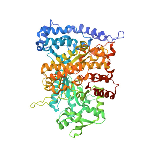
The high fidelity of DNA replication and repair is attributable, in part, to the allosteric regulation of ribonucleotide reductases (RNRs) that maintains proper deoxynucleotide pool sizes and ratios in vivo. In class Ia RNRs, ATP (stimulatory) and dATP (inhibitory) regulate activity by binding to the ATP-cone domain at the N terminus of the large α subunit and altering the enzyme's quaternary structure. Class Ib RNRs, in contrast, have a partial cone domain and have generally been found to be insensitive to dATP inhibition. An exception is the Bacillus subtilis Ib RNR, which we recently reported to be inhibited by physiological concentrations of dATP. Here, we demonstrate that the α subunit of this RNR contains tightly bound deoxyadenosine 5'-monophosphate (dAMP) in its N-terminal domain and that dATP inhibition of CDP reduction is enhanced by its presence. X-ray crystallography reveals a previously unobserved (noncanonical) α 2 dimer with its entire interface composed of the partial N-terminal cone domains, each binding a dAMP molecule. Using small-angle X-ray scattering (SAXS), we show that this noncanonical α 2 dimer is the predominant form of the dAMP-bound α in solution and further show that addition of dATP leads to the formation of larger oligomers. Based on this information, we propose a model to describe the mechanism by which the noncanonical α 2 inhibits the activity of the B. subtilis Ib RNR in a dATP- and dAMP-dependent manner.
- Department of Chemistry, Massachusetts Institute of Technology, Cambridge, MA 02139.
Organizational Affiliation: