Structural Basis of the Molecular Switch between Phosphatase and Mutase Functions of Human Phosphomannomutase 1 under Ischemic Conditions.
Ji, T., Zhang, C., Zheng, L., Dunaway-Mariano, D., Allen, K.N.(2018) Biochemistry 57: 3480-3492
- PubMed: 29695157 Search on PubMedSearch on PubMed Central
- DOI: https://doi.org/10.1021/acs.biochem.8b00223
- Primary Citation Related Structures:
6CFR, 6CFS, 6CFT, 6CFU, 6CFV - PubMed Abstract:
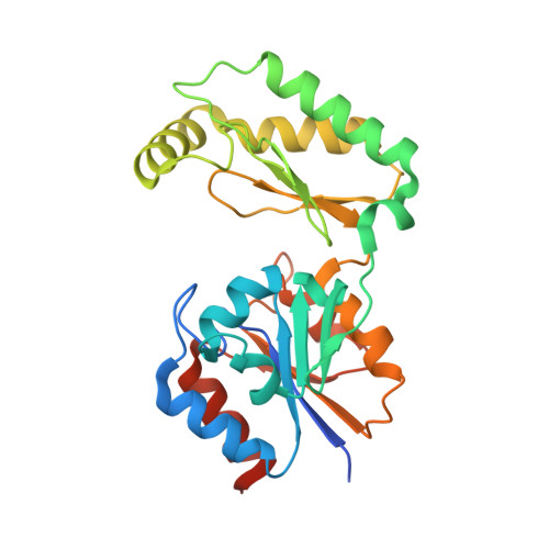
The human phosphomannomutases PMM1 and PMM2 catalyze the interconversion of hexose 6-phosphates and hexose 1-phosphates. The two isoforms share 66% sequence identity and have kinetic properties similar to those of mutases in vitro but differ in their functional roles in vivo. Though the physiological role of PMM2 is catalysis of the mutase reaction that provides the mannose 1-phosphate (Man-1-P) essential for protein glycosylation, PMM1 is thought to provide a phosphohydrolase activity in the presence of inosine monophosphate (IMP), converting glucose 1,6-bisphosphate (Glu-1,6-P 2 ) to glucose 6-phosphate (Glu-6-P), rescuing glycolysis during brain ischemia. To uncover the structural basis of how IMP binding converts PMM1 from a mutase to a phosphatase, the 1.93 Å resolution structure of PMM1 complexed with IMP was determined. The structure reveals IMP bound at the substrate recruitment site, thus inhibiting the mutase activity while simultaneously activating a phosphatase activity (IMP K act = 1.5 μM) resulting from the hydrolysis of the phospho-enzyme. The bound structure and site-directed mutagenesis confirm that the long-range electrostatic interactions provided by Arg180 and Arg183 conserved in PMM1 are the major contributors to IMP binding, and their oblation removes phosphatase but not mutase activity. These residues are not present in the PMM2 isoform, which consequently lacks significant phosphatase activity in the presence of IMP. T 2 relaxation nuclear magnetic resonance and small angle X-ray scattering together support the hypothesis that binding of IMP to PMM1 favors an enzyme conformation that is catalytically competent for water attack at the phosphoaspartyl intermediate. Such a mechanism may be generalizable to other enzymes that act through covalent intermediates.
- Department of Chemistry , Boston University , Boston , Massachusetts 02215 , United States.
Organizational Affiliation: