6CFF

Stimulator of Interferon Genes Human


Experimental Data Snapshot

  • Method: X-RAY DIFFRACTION
  • Resolution: 2.40 Å
  • R-Value Free: 0.249 
  • R-Value Work: 0.193 
  • R-Value Observed: 0.198 

wwPDB Validation   3D Report Full Report


Ligand Structure Quality Assessment 


This is version 1.4 of the entry. See complete history


Literature

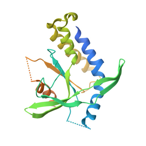
STING Polymer Structure Reveals Mechanisms for Activation, Hyperactivation, and Inhibition.

Ergun, S.L.Fernandez, D.Weiss, T.M.Li, L.

(2019) Cell 178: 290-301.e10

  • DOI: https://doi.org/10.1016/j.cell.2019.05.036
  • Primary Citation of Related Structures:  
    6CFF, 6CY7, 6DNK

  • PubMed Abstract: 

    How the central innate immune protein, STING, is activated by its ligands remains unknown. Here, using structural biology and biochemistry, we report that the metazoan second messenger 2'3'-cGAMP induces closing of the human STING homodimer and release of the STING C-terminal tail, which exposes a polymerization interface on the STING dimer and leads to the formation of disulfide-linked polymers via cysteine residue 148. Disease-causing hyperactive STING mutations either flank C148 and depend on disulfide formation or reside in the C-terminal tail binding site and cause constitutive C-terminal tail release and polymerization. Finally, bacterial cyclic-di-GMP induces an alternative active STING conformation, activates STING in a cooperative manner, and acts as a partial antagonist of 2'3'-cGAMP signaling. Our insights explain the tight control of STING signaling given varying background activation signals and provide a therapeutic hypothesis for autoimmune syndrome treatment.


  • Organizational Affiliation

    Department of Biochemistry, Stanford School of Medicine, Stanford University, Stanford, CA 94305, USA.


Macromolecules
Find similar proteins by:  (by identity cutoff)  |  3D Structure
Entity ID: 1
MoleculeChains Sequence LengthOrganismDetailsImage
Stimulator of interferon genes protein241Homo sapiensMutation(s): 1 
Gene Names: TMEM173ERISMITASTING
UniProt & NIH Common Fund Data Resources
Find proteins for Q86WV6 (Homo sapiens)
Explore Q86WV6 
Go to UniProtKB:  Q86WV6
PHAROS:  Q86WV6
GTEx:  ENSG00000184584 
Entity Groups  
Sequence Clusters30% Identity50% Identity70% Identity90% Identity95% Identity100% Identity
UniProt GroupQ86WV6
Sequence Annotations
Expand
  • Reference Sequence
Small Molecules
Ligands 1 Unique
IDChains Name / Formula / InChI Key2D Diagram3D Interactions
2BA
Query on 2BA

Download Ideal Coordinates CCD File 
B [auth A](2R,3R,3aS,5R,7aR,9R,10R,10aS,12R,14aR)-2,9-bis(6-amino-9H-purin-9-yl)octahydro-2H,7H-difuro[3,2-d:3',2'-j][1,3,7,9,2,8 ]tetraoxadiphosphacyclododecine-3,5,10,12-tetrol 5,12-dioxide
C20 H24 N10 O12 P2
PDXMFTWFFKBFIN-XPWFQUROSA-N
Experimental Data & Validation

Experimental Data

  • Method: X-RAY DIFFRACTION
  • Resolution: 2.40 Å
  • R-Value Free: 0.249 
  • R-Value Work: 0.193 
  • R-Value Observed: 0.198 
  • Space Group: P 41 21 2
Unit Cell:
Length ( Å )Angle ( ˚ )
a = 110.93α = 90
b = 110.93β = 90
c = 36.03γ = 90
Software Package:
Software NamePurpose
REFMACrefinement
XDSdata reduction
SCALAdata scaling
PHASERphasing

Structure Validation

View Full Validation Report


Ligand Structure Quality Assessment 


Entry History 

Deposition Data

Revision History  (Full details and data files)

  • Version 1.0: 2019-03-13
    Type: Initial release
  • Version 1.1: 2019-07-17
    Changes: Data collection, Database references
  • Version 1.2: 2019-07-24
    Changes: Data collection, Database references
  • Version 1.3: 2019-08-07
    Changes: Data collection, Database references
  • Version 1.4: 2024-03-13
    Changes: Data collection, Database references, Derived calculations, Structure summary