Rationally designed carbohydrate-occluded epitopes elicit HIV-1 Env-specific antibodies.
Zhu, C., Dukhovlinova, E., Council, O., Ping, L., Faison, E.M., Prabhu, S.S., Potter, E.L., Upton, S.L., Yin, G., Fay, J.M., Kincer, L.P., Spielvogel, E., Campbell, S.L., Benhabbour, S.R., Ke, H., Swanstrom, R., Dokholyan, N.V.(2019) Nat Commun 10: 948-948
- PubMed: 30814513 Search on PubMedSearch on PubMed Central
- DOI: https://doi.org/10.1038/s41467-019-08876-w
- Primary Citation Related Structures:
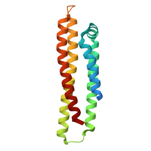
6CBU, 6CFE - PubMed Abstract:
An array of carbohydrates masks the HIV-1 surface protein Env, contributing to the evasion of humoral immunity. In most HIV-1 isolates 'glycan holes' occur due to natural sequence variation, potentially revealing the underlying protein surface to the immune system. Here we computationally design epitopes that mimic such surface features (carbohydrate-occluded neutralization epitopes or CONE) of Env through 'epitope transplantation', in which the target region is presented on a carrier protein scaffold with preserved structural properties. Scaffolds displaying the four CONEs are examined for structure and immunogenicity. Crystal structures of two designed proteins reflect the computational models and accurately mimic the native conformations of CONEs. The sera from rabbits immunized with several CONE immunogens display Env binding activity. Our method determines essential structural elements for targets of protective antibodies. The ability to design immunogens with high mimicry to viral proteins also makes possible the exploration of new templates for vaccine development.
- Department of Biochemistry and Biophysics, University of North Carolina at Chapel Hill, Chapel Hill, NC, 27599, USA.
Organizational Affiliation: