


Crystal Structure of the Human CAMKK1A in complex with GSK650394
Santiago, A.S., Counago, R.M., Righetto, G.L., Ramos, P.Z., Silva, P.N.B., Drewry, D., Elkins, J.M., Massirer, K.B., Arruda, P., Edwards, A.M.To be published.
Experimental Data Snapshot
Starting Model: experimental
View more details
Entity ID: 1 | |||||
|---|---|---|---|---|---|
| Molecule | Chains | Sequence Length | Organism | Details | Image |
| Calcium/calmodulin-dependent protein kinase kinase 1 | 290 | Homo sapiens | Mutation(s): 0 Gene Names: CAMKK1, CAMKKA EC: 2.7.11.17 | 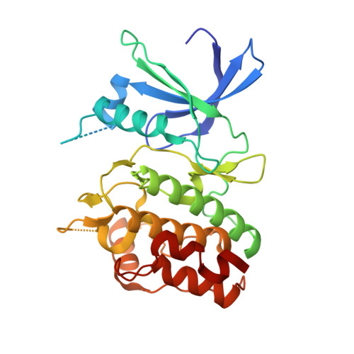 | |
UniProt & NIH Common Fund Data Resources | |||||
PHAROS: Q8N5S9 GTEx: ENSG00000004660 | |||||
Entity Groups | |||||
| Sequence Clusters | 30% Identity50% Identity70% Identity90% Identity95% Identity100% Identity | ||||
| UniProt Group | Q8N5S9 | ||||
Sequence AnnotationsExpand | |||||
Reference Sequence | |||||
| Ligands 2 Unique | |||||
|---|---|---|---|---|---|
| ID | Chains | Name / Formula / InChI Key | 2D Diagram | 3D Interactions | |
| DXV Download:Ideal Coordinates CCD File | E [auth A], G [auth B], I [auth C], K [auth D] | 2-cyclopentyl-4-(5-phenyl-1H-pyrrolo[2,3-b]pyridin-3-yl)benzoic acid C25 H22 N2 O2 WVSBGSNVCDAMCF-UHFFFAOYSA-N |  | ||
| CL Download:Ideal Coordinates CCD File | F [auth A], H [auth B], J [auth C], L [auth D] | CHLORIDE ION Cl VEXZGXHMUGYJMC-UHFFFAOYSA-M |  | ||
| Length ( Å ) | Angle ( ˚ ) |
|---|---|
| a = 44.378 | α = 90 |
| b = 96.042 | β = 90.03 |
| c = 141.26 | γ = 90 |
| Software Name | Purpose |
|---|---|
| REFMAC | refinement |
| XDS | data reduction |
| Aimless | data scaling |
| PHASER | phasing |
| Funding Organization | Location | Grant Number |
|---|---|---|
| Sao Paulo Research Foundation (FAPESP) | Brazil | 13/50724-5 |


