


Crystal structure of LpxC from Pseudomonas aeruginosa in complex with PT805
Phan, J.N., Delker, S.L., Lorimer, D.D., Horanyi, P.S., Edwards, T.E.To be published.
Experimental Data Snapshot
Starting Model: experimental
View more details
Entity ID: 1 | |||||
|---|---|---|---|---|---|
| Molecule | Chains | Sequence Length | Organism | Details | Image |
| UDP-3-O-acyl-N-acetylglucosamine deacetylase | 304 | Pseudomonas aeruginosa | Mutation(s): 0 Gene Names: lpxC, PLES_47851 EC: 3.5.1.108 | 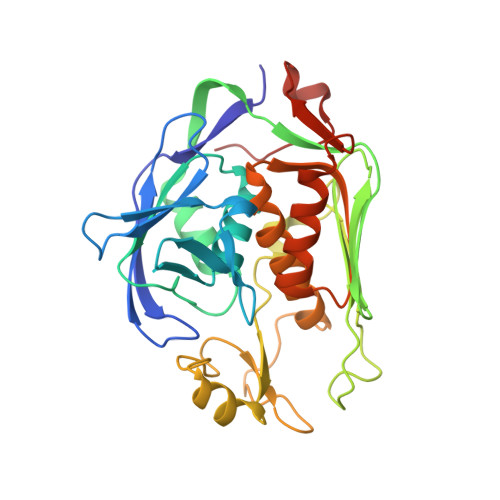 | |
UniProt | |||||
Entity Groups | |||||
| Sequence Clusters | 30% Identity50% Identity70% Identity90% Identity95% Identity100% Identity | ||||
| UniProt Group | B7UZI4 | ||||
Sequence AnnotationsExpand | |||||
Reference Sequence | |||||
| Ligands 4 Unique | |||||
|---|---|---|---|---|---|
| ID | Chains | Name / Formula / InChI Key | 2D Diagram | 3D Interactions | |
| EUY (Subject of Investigation/LOI) Download:Ideal Coordinates CCD File | B [auth A], C [auth A] | (2R)-N-hydroxy-2-methyl-2-(methylsulfonyl)-4-{2-oxo-4-[4-(2H-1,2,3-triazol-2-yl)phenyl]pyridin-1(2H)-yl}butanamide C19 H21 N5 O5 S QSXIGYJNQBFDCD-LJQANCHMSA-N |  | ||
| ZN Download:Ideal Coordinates CCD File | D [auth A], E [auth A] | ZINC ION Zn PTFCDOFLOPIGGS-UHFFFAOYSA-N |  | ||
| EDO Download:Ideal Coordinates CCD File | G [auth A] H [auth A] I [auth A] J [auth A] K [auth A] | 1,2-ETHANEDIOL C2 H6 O2 LYCAIKOWRPUZTN-UHFFFAOYSA-N |  | ||
| CL Download:Ideal Coordinates CCD File | F [auth A] | CHLORIDE ION Cl VEXZGXHMUGYJMC-UHFFFAOYSA-M |  | ||
| Length ( Å ) | Angle ( ˚ ) |
|---|---|
| a = 35.76 | α = 111.26 |
| b = 47.47 | β = 109.17 |
| c = 48.23 | γ = 98 |
| Software Name | Purpose |
|---|---|
| XDS | data reduction |
| XSCALE | data scaling |
| PHENIX | refinement |
| PDB_EXTRACT | data extraction |
| Coot | model building |


