Effects of rigidity on the selectivity of protein kinase inhibitors.
Assadieskandar, A., Yu, C., Maisonneuve, P., Liu, X., Chen, Y.C., Prakash, G.K.S., Kurinov, I., Sicheri, F., Zhang, C.(2018) Eur J Med Chem 146: 519-528
- PubMed: 29407977 Search on PubMedSearch on PubMed Central
- DOI: https://doi.org/10.1016/j.ejmech.2018.01.053
- Primary Citation Related Structures:
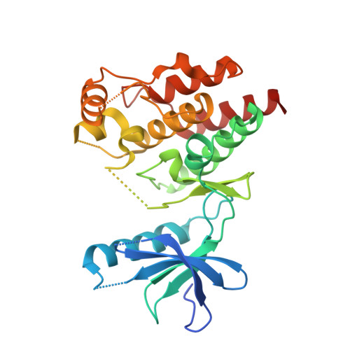
6CAD - PubMed Abstract:
Established strategies for discovering selective kinase inhibitors are target-centric as they often target certain structural or reactive features in the target kinase. In the absence of such prominent features, there is a lack of general methods for discovering selective inhibitors. Here we describe a new strategy that exploits conformational flexibility of kinases for achieving selective kinase inhibition. Through ring closure, we designed and synthesized a panel of isoquinoline-containing compounds as rigidified analogs of an amidophenyl-containing parent compound. These analogs potently inhibit kinases including Abl and BRAF but have diminished inhibition against some other kinases compared to the parent compound. Sequence analysis reveals that many of the kinases that are potently inhibited by the isoquonoline-containing compounds contain a long insertion within their catalytic domains. A crystal structure of one rigid compound bound to BRAF confirmed its binding mode. Our findings highlight the potential of a novel strategy of rigidification for improving the selectivity of kinase inhibitors.
- Loker Hydrocarbon Research Institute & Department of Chemistry, University of Southern California, University Park, Los Angeles, CA 90089, USA.
Organizational Affiliation: