New fluorescence-based high-throughput screening assay for small molecule inhibitors of tyrosyl-DNA phosphodiesterase 2 (TDP2).
Ribeiro, C.J.A., Kankanala, J., Shi, K., Kurahashi, K., Kiselev, E., Ravji, A., Pommier, Y., Aihara, H., Wang, Z.(2018) Eur J Pharm Sci 118: 67-79
- PubMed: 29574079 Search on PubMedSearch on PubMed Central
- DOI: https://doi.org/10.1016/j.ejps.2018.03.021
- Primary Citation Related Structures:
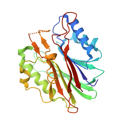
6CA4 - PubMed Abstract:
Tyrosyl-DNA phosphodiesterase 2 (TDP2) repairs topoisomerase II (TOP2) mediated DNA damages and causes resistance to TOP2-targeted cancer therapy. Inhibiting TDP2 could sensitize cancer cells toward TOP2 inhibitors. However, potent TDP2 inhibitors with favorable physicochemical properties are not yet reported. Therefore, there is a need to search for novel molecular scaffolds capable of inhibiting TDP2. We report herein a new simple, robust, homogenous mix-and-read fluorescence biochemical assay based using humanized zebrafish TDP2 (14M_zTDP2), which provides biochemical and molecular structure basis for TDP2 inhibitor discovery. The assay was validated by screening a preselected library of 1600 compounds (Z' ≥ 0.72) in a 384-well format, and by running in parallel gel-based assays with fluorescent DNA substrates. This library was curated via virtual high throughput screening (vHTS) of 460,000 compounds from Chembridge Library, using the crystal structure of the novel surrogate protein 14M_zTDP2. From this primary screening, we selected the best 32 compounds (2% of the library) to further assess their TDP2 inhibition potential, leading to the IC 50 determination of 10 compounds. Based on the dose-response curve profile, pan-assay interference compounds (PAINS) structure identification, physicochemical properties and efficiency parameters, two hit compounds, 11a and 19a, were tested using a novel secondary fluorescence gel-based assay. Preliminary structure-activity relationship (SAR) studies identified guanidine derivative 12a as an improved hit with a 6.4-fold increase in potency over the original HTS hit 11a. This study highlights the importance of the development of combination approaches (biochemistry, crystallography and high throughput screening) for the discovery of TDP2 inhibitors.
- Center for Drug Design, Academic Health Center, University of Minnesota, Minneapolis, MN 55455, United States.
Organizational Affiliation: